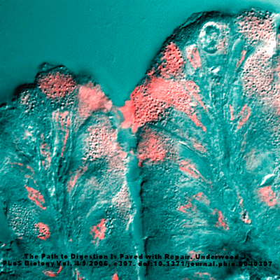
The nematode Heligmosomoides polygyrus, which was used in this study, seen into an optical microscope. Taken from the digestive tractus of a rodent.
Helminths, or gut worms, are known to be powerful suppressants of the immune system. In fact, this is the basis for using helminth therapy for various autoimmune conditions, such as IBD. Still though, the mechanisms for helminth immunosuppression is unknown. There have been some studies that suggest the worms are secreting molecules that have this anti-inflammatory effect, but this may not tell the whole story. Researchers from Switzerland hypothesized that because helminths and our gut bacteria evolved together, it was likely that the helminths were modulating the bacterial gut microbiome, and that this modulation was anti-inflammatory. They tested and published results that support this idea in the latest issue of Cell Immunity.
The scientists started by showing the efficacy of a mouse helminth, Heligmosomoides polygyrus bakeri (Hpb), in reducing inflammation in mouse models of asthma. The scientists infected mice with the parasite and exposed those mice, along with non-infected control mice, to dust mites in order to elicit and immune response. The scientists observed that the Hpb mice had much lower circulating levels of specific cytokines and immune cells after exposure to dust mites than the controls. Next, the scientist gave the Hpb infected mice antibiotics, which eliminated the gut bacteria but left the helminths intact. They then exposed these mice and control mice to dust mites to elicit the immune response. Interestingly, while the helminths alone did decrease the levels of some inflammatory molecules and cells, inflammation still occurred, similar to what was observed in controls. This meant that the gut bacteria play a role in modulating the helminthic immune suppression. In order to validate these findings, the scientists then performed fecal microbiota transplants from control mice or helminth infected mice into germ free mice (with no worms). After, the challenged these mice with house dust mites and discovered that the gut bacteria alone created an immune suppression in the mice, even in the absence of the worms.
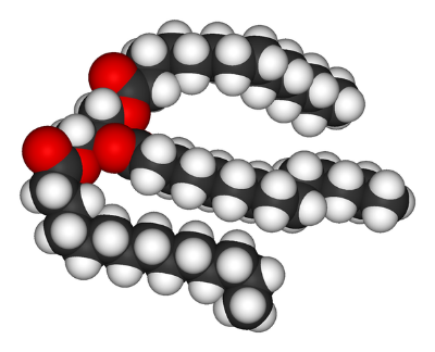
The researchers attempted to identify which bacteria may be causing this immune suppression, and measured the microbiomes of the mice. They noted that higher levels of Clostridiales occurred in the Hpb mice. They then measured the levels of short chain fatty acids (SCFAs) in the mice’s guts, because Clostridiales are known to produce SCFAs. They noticed that higher levels of SCFAs, which have previously been linked to immune suppression, did occur in higher levels in mice with Hpb compared to controls. The scientists then studied this connection between worm infection and increase in SCFAs in pigs and humans. Remarkably, the increase in SCFAs in helminth-infected subjects compared to controls was observed across species, suggesting the immune suppressing helminth phenomenon is extensible to many mammals. The researchers even investigated possible mechanisms for why SCFAs were able to suppress the immune system. They discovered the SCFAs were binding specific receptors that modulate T-cells, and more depth on this issue can be found by reading the paper.
This study is quite important as it shows that helminths in combination with the bacterial microbiome are important to immune suppression. This suggests that future therapeutics that may take advantage of helminth-derived molecules may not be as effective. It does, however, support helminth therapy as an immune suppressant. However, helminths are also very dangerous and can lead to various diseases. So, while clinical trials that use helminths are underway, there are still no approved uses for worms.