sialic acid

Bacteria from infants’ microbiome metabolize breast milk differently.

Human milk oligosaccharides (HMOs) are a diverse group of carbohydrates found in breastmilk.  Because the HMOs can’t be used by the infant directly for energy, scientists believe their purpose is to stimulate the development of a healthy gut microbiome.  During the first year of life, an infant’s gut is dominated by Bifidobacteria, in particular B. infantis, and B. bifidum.  In a recent publication scientists measured the difference in HMO utilization between these bugs, and discovered they have very different and important strategies for HMO utilization. The results were published in Nature Scientific Reports.

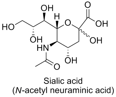
The scientists first isolated multiple strains of each species, B. infantis and B. bifidum, from the feces of newborn infants.  They then attempted to culture each strain alone in a mixture of HMOs from breastmilk, as well as the individual HMOs alone.  They learned that the each B. infantum strain could grow on pooled HMOs, but interestingly some of the B. bifidum strains could not grow alone on HMOs.  When the bacteria were cultured with mucins (containing sialic acid or fucose, as previously discussed on this blog) none of the B. infantis could grow, whereas most of the B. bifidum could.  This implies that B. infantis alone cannot utilize fucose or sialic acid, but rather needs the help of other bugs to break these down to utilize them.  After, the scientists looked at the regulation of different genes during the culturing experiments.  From these results they determined that B. infantum transports the HMOs inside the cell before breaking them down for energy.  B. bifidum, on the other hand, breaks the oligosaccharides down extracellularly before taking up smaller, simpler sugars. 

All together we see that there is a complex assemblage of bugs in the guts of infants that all rely on one another for energy and metabolism.  The breastmilk cocktail of HMOs itself is so complicated that it almost necessitates the interdependent communities to grow.  Overall, this creates a robust and resilient microbiome that prevents pathogens from taking hold and protects the infant during his or her most vulnerable years.

Please email blog@MicrobiomeInstitute.org for any comments, news, or ideas for new blog posts.

The views expressed in the blog are solely those of the author of the blog and not necessarily the American Microbiome Institute or any of our scientists, sponsors, donors, or affiliates.

Sialic acid may be key carbohydrate responsible for inflammation and dysbiosis in the gut

A surface mucous cell bordering on the stomach lumen secretes mucus (pink stain).

A surface mucous cell bordering on the stomach lumen secretes mucus (pink stain).

Our diet is full of various carbohydrates, composed of different monosaccharides and polysaccharides.  Many of these survive our own digestion and make it all the way to the colon where they modulate our microbiome.  Another source of saccharides for our gut bacteria is the mucous that we produce, which can be a rich source of fucose or sialic acid.  Sialic acid has been implicated in many inflammatory diseases, such as bacterial vaginosis.  Last week, researchers from Switzerland showed that sialic acid may play a critical role in colitis, at least in one colitis model commonly used in mice.  They published their results in Nature Communications.

One way to induce intestinal inflammation in mice is to feed them dextran sodium sulfate (DSS).  The reason this molecule causes colitis in this mice is unknown, but it is used in many models of the disease.  In order to understand the possible role of sialic acid in the colitis, scientists created mice that could not produce mucous with sialic acid.  They quickly realized that these mice were not as susceptible to the DSS-induced colitis as their normal counterparts.  After, they tested how various antibiotics affected colitis in the DSS—colitis mice and discovered that Escherichia coli abundance was directly associated with the severity of the disease. Putting these ideas together, they tested and discovered that E. coli used sialic acid as their main carbohydrate source in vitro.

Interestingly, the E. coli cannot actually access the sialic acid from mucins, but instead need other bacteria, such as Bacteroides vulgatus to cleave and release the sialic acid from the mucins in order to access it.  If sialic acid is indeed important to the human form of the disease there may be multiple approaches to combatting the disease.  First, by eliminating E. coli, and second by eliminating the free sialic acid.

Please email blog@MicrobiomeInstitute.org for any comments, news, or ideas for new blog posts.

The views expressed in the blog are solely those of the author of the blog and not necessarily the American Microbiome Institute or any of our scientists, sponsors, donors, or affiliates.

A new way in which some gut bacteria rely on their hosts’ mucous for energy

It is often understood that our gut bacteria live off of the foods we eat.  However, many gut bacteria can actually metabolize the mucous that protects the lining of our gut.  In fact, many bugs have the ability to digest a specific sugar that is attached to our mucins, called sialic acid.  Interestingly, some bacteria have the ability to cleave this sugar from the mucins, others have the ability to consume this sugar once it is released, and others have the ability to perform both of these tasks.  Last week, a new method for how gut bacteria can transform sialic acid was discovered, that some bacteria can actually transform sialic acid before cleaving it, and that this may be clinically relevant for Crohn’s disease and colitis.  The authors published their results in Nature Communications.

The authors were testing a common commensal bacteria, Ruminococcus gnavus, and noted that it had the ability to both cleave and consume sialic acid from gut mucins.  When they identified the metabolites from this process they discovered that the sialic acid was actually being converted to a different form by these bugs.  After further experimentation they realized that a type of enzyme, called an intramolecular trans sialidase, which had never before been observed in gut bacteria, was responsible.  The researchers then compared the genes from R. gnavus to other bugs common in the gut and noted that a full 11% of human gut commensals had this enzyme, and that these bacteria were overrepresented in people with IBD.  The authors think that the bugs who code for this enzyme have an inherent advantage over other gut microbiota because after they transform the sialic acid they can still use it for energy, whereas other bugs cannot, leaving the sugar all to themselves. 

The paper did not discuss specific mechanisms as to why these bugs may be overrepresented in Crohn’s and colitis.  They did however test a few molecules that inhibited the activity of the enzymes.  Perhaps if these enzymes or the responsible bugs are the cause of IBD, than these inhibitors could be used as therapeutics to combat the disease. 

Please email blog@MicrobiomeInstitute.org for any comments, news, or ideas for new blog posts.

The views expressed in the blog are solely those of the author of the blog and not necessarily the American Microbiome Institute or any of our scientists, sponsors, donors, or affiliates.

Breastmilk varies between mothers – affects microbiome of infant

Chemical structure of fucose molecule

Chemical structure of fucose molecule

We know that breastmilk is crucial to the development of a healthy infant’s microbiome.  It contains many oligosaccharides that cannot be digested by the infant, and whose primary purpose appears to be stimulating the growth of specific microbiome bugs.  There are, however, differences between new mothers’ milk.  For instance, some mothers cannot produce 2′-fucosylated oligosaccharides, which are oligosaccharides that have a fucose sugar on the end.  David Mills and his team at UC Davis recently investigated how the microbiomes of infants differed based on the presence or absence of fucosylated glycans in the milk that they drank.  They published their work in the journal Microbiome last week.

Forty four infants who were fed breast milk had their microbiomes measured throughout the first 120 days of their lives.  Thirty two of these infants were fed milk from woman with fucosylation ability (secretors), and twelve were from women without the fucosylation ability (non-secretors).  When the researchers investigated the contents of the milk they found that it varied in many ways, besides fucosylation.  For example, those women that did not fucosylate appeared instead to increase their monosaccharide sialylation, a sugar that has been linked to C. difficile infection.  When the scientists compared the infants’ microbiomes in the two groups they discovered that secretor-fed infants achieved higher levels of Bifidobacteria and Bacteroides, and achieved these levels more quickly than non-secretor-fed infants.  Instead, the non-secretor-fed infants had relatively higher levels of Enterobacteria, Clostridia, and Streptococci.

These differences may be important to the infants’ developments.  For example, Bifidobacteria in the gut is associated with lower gut permeability and less inflammation.  Also, Bifidobacteria and Bacteroides are large contributors to the production of short chain fatty acids and lactate, which have each been associated with gut health time and time again.  A full 20% of the U.S. population is non-secretors, and it would be interesting to see if any epidemiologically significant differences exist between the two groups into adulthood.  In either case, in the future it may be worth considering supplementing infant milk with fucosylated oligosaccharides if the lack of fucosylation does turn out to be detrimental to the baby.

Please email blog@MicrobiomeInstitute.org for any comments, news, or ideas for new blog posts.

The views expressed in the blog are solely those of the author of the blog and not necessarily the American Microbiome Institute or any of our scientists, sponsors, donors, or affiliates.