Escherichia coli

Proton pump inhibitors affect the microbiome

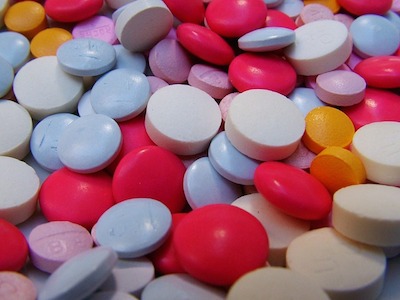
Proton pump inhibitors (PPI) are used to reduce gastric acid production in individuals’ guts and are prescribed to treat ulcers, gastroesophogeal reflux disease (GERD), and other conditions associated with acid production. It is one of the most commonly used drugs in the world. We know (and have written about) that PPIs are associated with increased intestinal infections, specifically Clostridium difficile, and the gut microbiome plays an important role in infections of the intestine. A recent study looked at the influence that PPIs had on the gut microbiome.

The team of researchers studied the gut microbiome of 1815 individuals. They looked at PPI users vs non-users. Of those sampled, 215 of them were taking a PPI at the time that a sample was taken. It was found that those taking the PPIs had lower microbial diversity compared to those not taking PPIs. They also found that bacteria usually found in the mouth was over-represented in the fecal samples of those taking PPIs, including those in the Rothia genus. They also observed an increase in EnterococcusStreptococcus, Staphylococcus, and Escherichia coli, a potentially pathogenic bacterium.

PPI usage effects are more prominent than those of most other drugs, including antibiotics. The results of this study are consistent with a less healthy microbiome and allow us to better understand why PPIs may lead to an increase of susceptibility to intestinal infections like C. diff.

Please email blog@MicrobiomeInstitute.org for any comments, news, or ideas for new blog posts.

The views expressed in the blog are solely those of the author of the blog and not necessarily the American Microbiome Institute or any of our scientists, sponsors, donors, or affiliates.

The microbiome can protect against metabolic dysregulation brought on by disease

Pathogenic infections can lead to metabolic alterations that result in maladies such as cachexia, or muscle wasting.  Antibiotics can be prescribed to treat a variety of intestinal diseases or inflammatory conditions, but these agents can also disrupt the natural microbiome ecology that could perhaps provide benefits to protecting against metabolic dysregulations.  On top of this, harnessing components of the microbiome with respect disease tolerance is an avenue under continuous exploration.  Within this contextual framework, researchers from The Salk Institute for Biological Studies in La Jolla, CA investigated to see whether components of the microbiome could have a protective effect on metabolic dysregulation brought on by gut trauma and/or infection.   

To initiate the investigation, the researchers used an induced-injury model known as the dextran sulfate sodium (DSS) intestinal injury model to create symptoms associated with inflammatory bowel disease/Crohn’s disease.  DSS was applied to mice in two cohorts procured from two distinct laboratories (Jackson labs [Jax] and UC Berkeley lab [CB]).  This treatment was administered to C57 mice followed by an administration of an antibiotic cocktail of ampicillin, vancomycin, neomycin, and metronidazole (AVNM) to provide remedy for the injury.  The AVNM cocktail had no impact on the severity of DSS in mice procured from Jackson labs, whereas mice procured from UC Berkeley colonies demonstrated significantly less muscle wasting.  This observation led to the hypothesis that microbiota composition differences between both cohorts of mice drove this observation. 

After examining cecal content from AVNM-CB and AVNM-Jax mice, it was determined that the CB mice had a higher composition of E. coli compared to the Jax mice.  Building on the original supposition, the researchers then administered E. coli to Jax mice, and upon DSS administration, they demonstrated significantly less wasting pathology as compared to the vehicle control groups (i.e., DSS treatment without being administered E. coli).  The researchers further investigated whether E. coli had a protective effect in response to infectious microbes in addition to induced-DSS injury, and Jax mice were infected with Salmonella Typhimurium or Burkholderia thailandensis.  There was no significant difference in alterations in host metabolism, caloric uptake, or inflammation between E. coli-administered groups and controls.  However, the E. coli group demonstrated increased signaling in the insulin-like growth factor 1/phosphatidylinositol 3-kinase/AKT pathway in skeletal muscle, a pathway implicated in the prevention of muscle wasting.  This finding effectively provides mechanistic evidence of protecting against muscle wasting. 

Together, these findings provide additional evidence that support the microbiome’s role in tempering inflammatory disease or injury.  Further delineation of molecular pathways associated with these maladies will advance our understanding and treatment of disease.   

Please email blog@MicrobiomeInstitute.org for any comments, news, or ideas for new blog posts.

The views expressed in the blog are solely those of the author of the blog and not necessarily the American Microbiome Institute or any of our scientists, sponsors, donors, or affiliates.

New study shows how E. coli and B. theta grow in the gut mucus

The mucosal membrane continues to be one of the most intriguing and vexing components of the gut microbiome.  It is the interface between the body and the environment, it is inhabited many bacteria, and it is a nutritional source that shapes the populations in the gut.  There is still very little known about the specific interactions between gut mucous and bacteria, but this critical system is rapidly being studied.  In the most recent advance, scientists from Switzerland and Germany examined two very different gut bacteria that fill different mucosal niches. They published their results in the journal Nature Communications.  The two bacteria they studied were Bacteroides thetaiotaomicron (B. theta) and Escherichia coliB. theta is a slow growing bacteria that has high metabolic flexibility that is capable of directly using gut mucins as an energy source.  E. coli is a fast growing bacteria that is much more limited in its metabolism and can’t directly use the carbohydrates in the gut, but can take hold and rapidly proliferate after a course of antibiotics. 

The researchers meticulously researched gnotobiotic mice and made many discoveries about bacteria in their mucous.  First, they discovered that the mucosal microbiome varies across its thickness, and is sterile closest to the intestines, but rich in life closest to the lumen.  In addition, they noted that the luminal microbiome is distinct from the mucosal microbiome, even though the mucous is constantly being shed into the lumen.  To this end, they confirmed that with regards to E. coli, these bugs replicate faster than they are shed (in about 3 hours in the mucous but 8 hours in the lumen), and that their persistence is due to replication rather than uptake from the lumen.  How though, can E. coli thrive with their limited ability to break down mucins?  The scientists learned that they likely metabolize iron, in addition to atypical carbon sources such as fatty acids and glycerol.  B. theta, on the other hand, has a huge repertoire of genes to break down mucins.  They do, though, have the ability to leave the mucins and form biofilms on bits of food, such as fiber, that pass through the lumen, and this is one way they travel through the gut.  Regardless of whether they are in the lumen or the mucins they proliferate at the same rate.

Each of these bacteria occupy different niches in the gut, and each is important to our health.  The discovery that E. coli can use iron for metabolism is particularly interesting, as chemotrophy is not normally considered as important in the body, and may be important to iron regulation.  As more research is published the mucous appears to be ‘where the rubber meets the road’ in the microbiome, and new discoveries in this area will be crucial to our overall understanding of the microbiome’s interaction with the body.

Please email blog@MicrobiomeInstitute.org for any comments, news, or ideas for new blog posts.

The views expressed in the blog are solely those of the author of the blog and not necessarily the American Microbiome Institute or any of our scientists, sponsors, donors, or affiliates.

Infants’ saliva may react with breast milk to modulate their microbiomes

Breastmilk is critically important to developing a healthy infant gut microbiome.  The combination of oligosaccharides found in breastmilk are not found in any other individual food, and are intended to cultivate healthy bacteria in the gut.  Besides breast milk, really the only other fluid an infant consumes is his or her own saliva, but thus far not much is known about the role this saliva plays in culturing the proper microbiota.  A team of researchers from Australia recently studied how a mother’s breastmilk directly interacts with her infant’s saliva.  They discovered that when combined, saliva and breast milk produce specific molecules that inhibit the growth of some bacteria, but support the growth of others. They published their results in the journal PLoS ONE.

The researchers measured the molecular components of saliva in 77 adults and 60 infants.  They noticed some stark differences between the two types of saliva, including markedly higher levels of salivary hypoxanthine and xanthine.  Hypoxanthine and xanthine are both substrates for a protein called xanthine oxidase (XO), which reacts with them to form hydrogen peroxide (H2O2).  One of the places XO is predominantly found is in human breast milk, which led the researchers to hypothesize that xanthine and hypoxanthine in infant saliva reacts with XO in breastmilk to form H2O2.  Hydrogen peroxide is a reactive oxygen species (ROS) that can kill bacteria.  The scientists believe that infant saliva reacts with breast milk to form hydrogen peroxide at high enough levels to kill opportunistic pathogens, but allow others to grow.  In order to test their hypothesis, the researchers combined breast milk and infant saliva and attempted to culture the pathogen Staphylococcus aureus, along with gut commensal bacteria Lactobacillus plantarum, and Escherichia coli.  They found that the mixture created concentrations of hydrogen peroxide that killed the S. aureus but allowed the commensals to grow.

Overall this paper showed that infant saliva can combine with breast milk to form physiologically relevant concentrations of hydrogen peroxide.  The hydrogen peroxide may in fact select for the growth of specific bacteria in the mouth and gut, and lead to the development of a healthy microbiome.  Interestingly, pasteurized cow’s milk and infant formula did not contain XO, the enzyme necessary to create the hydrogen peroxide, adding another reason why there is no true substitute for breast milk.   

Please email blog@MicrobiomeInstitute.org for any comments, news, or ideas for new blog posts.

The views expressed in the blog are solely those of the author of the blog and not necessarily the American Microbiome Institute or any of our scientists, sponsors, donors, or affiliates.

Sialic acid may be key carbohydrate responsible for inflammation and dysbiosis in the gut

A surface mucous cell bordering on the stomach lumen secretes mucus (pink stain).

A surface mucous cell bordering on the stomach lumen secretes mucus (pink stain).

Our diet is full of various carbohydrates, composed of different monosaccharides and polysaccharides.  Many of these survive our own digestion and make it all the way to the colon where they modulate our microbiome.  Another source of saccharides for our gut bacteria is the mucous that we produce, which can be a rich source of fucose or sialic acid.  Sialic acid has been implicated in many inflammatory diseases, such as bacterial vaginosis.  Last week, researchers from Switzerland showed that sialic acid may play a critical role in colitis, at least in one colitis model commonly used in mice.  They published their results in Nature Communications.

One way to induce intestinal inflammation in mice is to feed them dextran sodium sulfate (DSS).  The reason this molecule causes colitis in this mice is unknown, but it is used in many models of the disease.  In order to understand the possible role of sialic acid in the colitis, scientists created mice that could not produce mucous with sialic acid.  They quickly realized that these mice were not as susceptible to the DSS-induced colitis as their normal counterparts.  After, they tested how various antibiotics affected colitis in the DSS—colitis mice and discovered that Escherichia coli abundance was directly associated with the severity of the disease. Putting these ideas together, they tested and discovered that E. coli used sialic acid as their main carbohydrate source in vitro.

Interestingly, the E. coli cannot actually access the sialic acid from mucins, but instead need other bacteria, such as Bacteroides vulgatus to cleave and release the sialic acid from the mucins in order to access it.  If sialic acid is indeed important to the human form of the disease there may be multiple approaches to combatting the disease.  First, by eliminating E. coli, and second by eliminating the free sialic acid.

Please email blog@MicrobiomeInstitute.org for any comments, news, or ideas for new blog posts.

The views expressed in the blog are solely those of the author of the blog and not necessarily the American Microbiome Institute or any of our scientists, sponsors, donors, or affiliates.

HIV vaccine failed because of interaction with microbiome

Scanning electron micrograph of HIV-1, colored green, on a lymphocyte.

Scanning electron micrograph of HIV-1, colored green, on a lymphocyte.

From 2009 to 2013, scientists at Duke University and the National Institute of Allergy and Infectious Diseases had been working on what looked like a promising cure to HIV. The study was terminated in 2013 because it was clear that the vaccine was not effective in protecting against HIV infection. An article recently published by Science Magazine gave insight into why the HIV vaccine unfortunately failed. Hint: it has something to do with the microbiome.

The vaccine was administered in the study to adult males in the form of an initial vaccine as well as a second booster vaccine. The HIV vaccine looked promising because it stimulated the body’s immune system to produce antibodies that recognize HIV. The unexpected result, however, was that these antibodies also recognized bacteria like Escherichia coli, a very important bacteria that lives in the human gut. It should be easy to see why this is a bad thing for the microbiome. Destroying important gut bacteria is very detrimental to humans, which we see over and over again here on the blog. Additionally, because the antibodies were reactive to bacteria as well as the HIV virus, it took away from the effectiveness of fighting HIV.

This study is very important in the search towards finding a cure to HIV, because it presented an unexpected obstacle that a lot can be learned from. Moving forward, questions are already being raised by the scientists such as, would this vaccine work for children if immunization was given to pregnant mothers? Perhaps the still-developing immune system would better be able to work with the vaccine. Only more research can prove whether the HIV vaccine is indeed still promising.  In addition, this may provide insights into the efficacy of vaccines for other diseases.  Perhaps the microbiome plays a large role in their effectiveness.  Vaccine research going forward should begin to take the microbiome into account.

Please email blog@MicrobiomeInstitute.org for any comments, news, or ideas for new blog posts.

The views expressed in the blog are solely those of the author of the blog and not necessarily the American Microbiome Institute or any of our scientists, sponsors, donors, or affiliates.