New study suggests S. aureus and skin dysbioses cause eczema

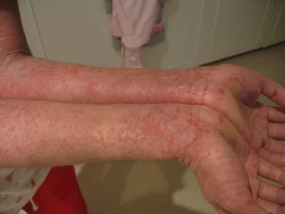
Atopic dermatitis, also known as eczema, is a skin inflammation and rash that has an enigmatic cause.  There are many genetic and environmental risk factors involved, but to date the exact triggers and mechanisms that cause this autoimmune response are unknown.  Importantly, a growing percentage of infants and toddlers are developing this disease, which itself is a known risk factor for other autoimmune diseases like asthma and allergies.  Discovering the cause of atopic dermatitis is an important endeavor, because it may lead to a cure for a number of other diseases.

Staphylococcus aureus has long been associated with atopic dermatitis.  It appears to occur at relatively high abundances in the areas of skin that are affected.  Then again, S. aureus is a one of the most common skin microbiome bacteria (it is ubiquitous around the world), and it has yet to be definitively connected to the disease.  In addition, mouse models for many skin diseases, including this one, do not exist or are insufficient, so controllably studying the atopic dermatitis is difficult.  Recently though, a team of scientists from Japan and the NIH developed a mouse model for atopic dermatitis, and made a new discovery that showed S. aureus can indeed drive skin inflammation.  They published their results in Cell immunity.

The scientists were studying how a specific genetic mutation in mice affected bones and hair follicles when they serendipitously realized that it was causing eczema in the mice after around three weeks.  When they investigated the skin microbiomes of these mice, as well as normal mice, they realized that right around the time that the eczema was appearing in the mice, these mice’s skin microbiomes drastically shifted.  First, a bacterium called Corynebacterium mastitidis emerged, followed by S. aureus a few weeks later, which was coincident with the presentation of the worst symptoms.  Of note, species of Corynebacteria are associated with eczema in humans, much like S. aureus.

Next, the researchers then performed a series of experiments by providing mice with antibiotics in an effort to combat the dysbiosis.  When newborn mice with the genetic modification were treated with antibiotics they never developed eczema at all.  Moreover, genetically modified mice that were in the midst of the rash that were treated with antibiotics had their eczema subside soon after.  In addition, genetically modified mice that were taken off of antibiotics had eczema emerge shortly thereafter.  Strikingly, in all of the above situations changes in the skin microbiome corresponded with the disease state: a lack of S. aureus and high diversity were associated with healthy skin, and the emergence of S. aureus and a lack of diversity were associated with the disease.  Finally, when S. aureus was inoculated onto the skin of genetically modified mice, they developed eczema rapidly.

The researchers performed a number of other experiments to try and tease out the mechanism by which S. aureus causes atopic dermatitis.  Their results show that it appears a combination of genetic and environmental factors that affect the skin may be important in defining an individual’s risk for the disease.  Regardless, it appears that S. aureus is a major culprit in causing eczema, so future therapies that eradicate the bacteria, or at least decrease its abundance should be considered.

Please email blog@MicrobiomeInstitute.org for any comments, news, or ideas for new blog posts.

The views expressed in the blog are solely those of the author of the blog and not necessarily the American Microbiome Institute or any of our scientists, sponsors, donors, or affiliates.

Immune cells are educated in the gut to not attack beneficial bacteria

The gastrointestinal tract is made up of trillions of bacteria that are largely ignored by the body’s immune system.  Why is it that the body’s immune system knows to ignore these beneficial bacteria that are so important for our ability to live a healthy life? The answer to this question could play an important role in understanding how to maintain a healthy gut and how to treat diseases. Scientists led by Gregory Sonnenberg at Weill Cornell Medical College may have answered this question in a study published last week in Science.

The researchers studied T cells, cells that are made in the thymus and are trained there to kill-off foreign microbes and other intruders that make their way into the human body. But why don’t these T cells attack helpful bacteria in the GI tract? They found that the T cells are again educated in the gut to not attack beneficial bacteria but when this education is disrupted, it can lead to disease.  For example, inflammatory bowel diseases like Crohn’s disease and ulcerative colitis occur when the immune system attacks the GI tract and bacteria in the GI tract.

In the thymus, T cells that could attack the body are destroyed before they are released into circulation. In the gut, a type of cell called innate lymphoid cells (ILCs) educate the T cells to not attack beneficial bacteria. These ILCs had previously been found to make a physical barrier between the bacteria in the gut and the immune system.

In mice, they found that ILCs attacked T cells that were destroying beneficial bacteria and when they prevented this attack by ILCs on the T cells, severe intestinal inflammation resulted. They also looked at intestinal biopsies of young patients with Crohn’s disease. In the biopsies they found that the ILCs lacked specific molecules that are important for educating the T cells not to attack the bacteria in the gut. They found that a decrease in this molecule correlated with an increase in pro-inflammatory cells in children with Crohn’s disease.

The authors state that it may be possible to get rid of these T cells that are causing the inflammation and by doing so you may be able to help treat the disease.  By restoring this molecule (Major Histocompatibility Complex class II) that is preventing the education of the T cells, pro-inflammatory T cells may be reduced resulting in reduced intestinal inflammation.


Please email blog@MicrobiomeInstitute.org for any comments, news, or ideas for new blog posts.

The views expressed in the blog are solely those of the author of the blog and not necessarily the American Microbiome Institute or any of our scientists, sponsors, donors, or affiliates.

Sex, body mass index, and dietary fiber correlated with microbiome composition

On last week’s podcast, we talked with Erica and Justin Sonnenburg about how the food we eat, and specifically dietary fiber, is important for “feeding” our microbiomes. All of the variables that influence microbiome composition are not fully understood, however research is continually being conducted to better understand what factors affect the microbiome.  To this end, a team of scientists from New York University School of Medicine set out to find how sex, body mass index (BMI), and dietary fiber intake impact the microbiome.

The scientists analyzed fecal samples from 82 individuals, 51 men and 31 women. They found that the women had different microbiome composition than the men, specifically a lower abundance of Bacteroidetes. They also found that BMI impacted microbiome diversity, specifically in women. Overweight and obese women had less diverse gut bacteria than normal weight women and women with a higher BMI also had less Bacteroidetes in their guts compared to the normal weight women.

The scientists also found that various sources of dietary fiber differentially impacted the microbiome of subjects.  Fiber intake from fruits and vegetables resulted in higher levels of Clostridia and fiber intake from beans was associated with greater abundance of Actinobacteria. It is possible that dietary fiber is influencing the microbiome by reducing gut transit time and lowering the pH. It is also possible that it is influencing systemic levels of estrogen, which could alter microbiome composition.

As the microbiome continues to be implicated in diseases, the ability to identify variables that affect the microbiome are important and can potentially be used for altering microbiota composition to prevent or possibly treat disease. 

Please email blog@MicrobiomeInstitute.org for any comments, news, or ideas for new blog posts.

The views expressed in the blog are solely those of the author of the blog and not necessarily the American Microbiome Institute or any of our scientists, sponsors, donors, or affiliates.

Significant bacterial diversity found in the microbiomes of remote Amerindians

A group of Yanomami people in Demini, Brazil © Fiona Watson/Survival

A group of Yanomami people in Demini, Brazil © Fiona Watson/Survival

The Yanomami Indians are a native people that reside in remote areas of the Amazon jungle in South America, where they live in a society devoid of westernization and modernization.  They were first contacted in the 1960s, and the Venezuelan government has since preserved isolation among these societies by preventing modern development from expanding into their lands.  From an anthropologic perspective, these people therefore represent a glimpse into the past of a hunter-gatherer subsistence living human society.  Capitalizing on this unique characteristic, researchers from several institutions led by AMI Scientific Advisory Board member Maria Gloria Dominguez Bello, set out to investigate the human microbiomes of these remote communities in a twofold manner.  Bacterial diversity of the Yanomami microbiota was characterized, concomitant to exploring bacterial and gene responses to antibiotics commonly utilized in clinics in Western societies. 

Thirty four Yanomami subjects between 4 and 50 years old were selected for analysis.  Forearm skin, oral mucosa, and fecal samples were collected for bacterial analysis and compared to subjects from the U.S., Guahibo Amerindian, and Malawian tribes (the latter two were selected for comparison, as their cultures are in transition to modernization).  E. coli cultures were examined and genomic libraries were created for further analysis of bacterial expression, functional diversity, and resistome (the collection of antibiotic resistant genes) gene expression in response to antibiotic treatment. 

The Yanomami people displayed extraordinary levels of bacterial diversity as compared to the U.S. subjects, Guahibo Amerindians, and Malawians.  Specifically, the Yanomami fecal samples were characterized by high expression of Prevotella and low expression of Bacteroide bacteria.  With respect to functional bacterial diversity, the Yanomami displayed higher fecal and skin functional diversity among the other Amerindian and U.S. subjects.  Furthermore, over-enrichment in bacteria that interact with pathways involved with protein and carbohydrate metabolism was observed in the Yanomami. 

23 antibiotics were tested on 131 E. coli strains isolated from 11 fecal samples, and sensitivity to all antibiotics was observed.  Interestingly, functional libraries created from these E. coli isolates displayed antibiotic resistant genes targeted against 8 of the 23 antibiotics.  This suggests that antibiotic resistance genes have been maintained despite the lack of apparent antibiotic selection pressure that is characteristic among Western societies. 

Results from this study indicate a greater scope of bacterial diversity in a defined group of people than ever reported before.  Furthermore, the investigation illuminated an important characteristic of the bacterial resistome, as the results indicated that resistant genes were present in a population that had not been exposed to any Westernization.  This suggests that some antibiotic resistant genes are archaic, dating back to pre-westernization times and also that westernization affects microbiome diversity.  Ultimately, this study employed a fascinating approach to investigating human microbiome divergence and evolution, in addition to providing more insight to host-species relationships with respect to pharmacologic therapies.  

Please email blog@MicrobiomeInstitute.org for any comments, news, or ideas for new blog posts.

The views expressed in the blog are solely those of the author of the blog and not necessarily the American Microbiome Institute or any of our scientists, sponsors, donors, or affiliates.

When you eat and what you eat may lead to obesity

Our bodies’ internal circadian clock may be profoundly important to our health, especially as it pertains to our metabolism.  Research has shown that people who have altered sleep cycles, like those who work the night shift, are at an increased risk for diabetes, obesity, and metabolic syndrome.  Researchers from the University of Chicago recently investigated how the microbiome may be involved in the complex relationship between disruptions to circadian rhythms and obesity.  They published their results in the journal Cell Host & Microbe.

The human circadian clock is regulated by a few organs in our bodies, including the brain, the liver, and is now evident from this study, the microbiome.  The researchers first measured gene regulation by the liver in germ free mice and normal mice.  They discovered that many of the genes that had daily rhythmic variations had their rhythms greatly affected by the presence and absence of bacteria in the gut.  They then subjected these mice to high fat and low fat diets and learned that, unsurprisingly, the high fat diet led to obesity in normal mice.  Surprisingly though, the high fat diet did not lead to obesity in germ-free mice.  Interestingly, many of the liver genes that were expressed rhythmically by the gut also had their rhythms affected by diet, with different genes having their expression altered depending on the diet.

The researchers then discovered that the populations of bacteria that comprise the microbiome also exhibited rhythmic variations throughout the day.  These variations did not necessarily relate to time of feeding either, as mice that were fed constantly throughout the day still experienced these variations.  Moreover, they realized that specific metabolic functions also changed rhythmically throughout the day, such as utilization of specific carbohydrates, and that a high fat diet would quell these rhythms.

The scientists then measured certain metabolites produced by the microbiome, such as short chained fatty acids (SCFAs), and saw these were also produced rhythmically throughout the day, which may be, but is not entirely, related to the differences in microbiome populations.  Metabolites rhythms were also affected by diet.  For example the high fat diet decreased SCFA rhythms.  The scientists then determined that these metabolites have a direct impact on the cycling of liver circadian genes.  This means that the microbiome metabolites and the human liver combine to contribute to our circadian clock.

The researchers go on to hypothesize that consuming a high fat diet disrupts our natural circadian rhythms, which leads to a lower metabolic state and results in obesity.  This hypothesis extends to the germ free mice which did not become obese regardless of diet; that is, they did not have a disrupted microbiome to alter their rhythms.  Ultimately, the healthiest and strongest circadian rhythms belonged to the normal mice eating normal food. 

We have written before about how jet lag can lead to microbiome changes that cause obesity.  This paper, in addition to the one described above show how our natural clock and the microbiome’s natural clock work in conjunction to regulate our metabolism.  Our circadian rhythms are not something which many people associate with the microbiome, but over time complex systems like this evolve.  While this paper may not make someone change his or her behavior, it may make him or her think twice before pulling an all-nighter or having that midnight snack. 

Please email blog@MicrobiomeInstitute.org for any comments, news, or ideas for new blog posts.

The views expressed in the blog are solely those of the author of the blog and not necessarily the American Microbiome Institute or any of our scientists, sponsors, donors, or affiliates.

How does a man’s seminal microbiome alter a woman’s vaginal microbiome?

There is very little research on the microbiome of semen.  We know that it is not sterile, and some scientists think that some of the bacteria found in semen may be involved in male fertility issues.  However, there is still a lot of research to be done in this area.  Even less is known about how the seminal microbiome influences the vaginal microbiome after sex.  Some research has suggested that specific sexual partners can cause bacterial vaginosis (BV), however the mechanisms for this are unclear.  It is suggested that perhaps the penile and seminal microbiome being transferred to the vagina during sex could cause this, although research has not confirmed these hypotheses.  Researchers from Estonia tried to answer these questions, and studied just how the vaginal and seminal microbiomes change before and after sex.  They published the results of their findings last week in Research in Microbiology

The scientists measured the seminal and vaginal microbiomes before and after sex for 23 couples who had sought help for infertility but were otherwise healthy.  They learned that the seminal microbiome, while containing much fewer bacteria, was actually more diverse than the vaginal microbiome.  Still though, each shared many of the same bacteria.  These included Lactobacillus, Veillonella, Streptococcus, Porphyromonas and Atopobium.  Interestingly, Gardnerella vaginalis, a bacterium highly implicated with BV, was found more frequently in women who had sex with men whose semen contained leukocytes, itself a phenotype associated with infertility.  While most of the women’s microbiomes did not shift after sexual intercourse, four of them did.  In these women a decrease in Lactobacillus occurred, and a decrease in Lactobacillus has also been highly implicated in BV.

While this study was preliminary, it marks some of the first research on the dynamics of the seminal and vaginal microbiome during sex.  The scientists suggest that the microbiome may be very important to fertility issues, and at the AMI we would not be surprised to learn that it is involved in at least some causes of infertility.  In the near future we will be devoting an entire podcast to the vaginal microbiome, and interviewing Jacques Ravel, a world leader in this field.  If you have any relavent questions and would like us to ask them on the podcast please call 518-945-8583 and leave your question on the voicemail.

Please email blog@MicrobiomeInstitute.org for any comments, news, or ideas for new blog posts.

The views expressed in the blog are solely those of the author of the blog and not necessarily the American Microbiome Institute or any of our scientists, sponsors, donors, or affiliates.