immune system

What happens if you give c-section babies a vaginal microbiome?

Babies born by cesarian section have greater likelihoods of autoimmune diseases during childhood and later in life.  They also have a gut microbiome that resembles their mother’s skin right after birth. On the other hand, babies that are born vaginally have a gut microbiome that resembles their mothers’ vaginas, and are at lower risk for asthma and allergies.  Given the importance of the microbiome on immune development, many scientists believe that there may be a link between mode of delivery, the initial infant gut microbiome, and normal immune development.

One possible method to ensure a baby that is born by c-section is initially colonized by his or her mother’s vaginal microbiome is to swab the mother’s vagina and transfer her microbiome to the baby immediately after birth.  Researchers from New York University performed this exact experiment, and measured the changes that occurred in the gut after this intervention.  They published their results in the journal Nature Medicine.

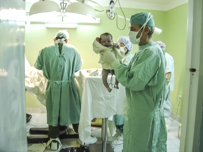
In the study, 18 women were split into 3 groups: 7 women gave birth naturally, 7 women gave birth by c-section, and 4 women gave birth by c-section but had their vaginal flora transferred to the babies.  This last group of women had their vaginas screened for pathogens shortly before birth.  After the c-section, and within 2 minutes after, gauze was rubbed in the new mothers’ vaginas and then rubbed all over babies’ mouths, faces, and bodies.  The babies’ skin and gut microbiomes were measured and compared to the other two groups.  As expected, the babies born vaginally had microbiomes that resembled their mothers’ vaginas, and the babies born by c-section had microbiomes that resembled their mothers’ skin.  Interestingly, the c-section babies that were inoculated with their mothers’ vaginal microbiomes, had a microbiome that closely resembled their mothers’ vaginas, even after 1 month.  In addition, there were no adverse consequences to the microbiome transfer.

This was a small proof of concept study that successfully showed a vaginal microbiome transfer to c-section babies could properly colonize a newly born infant.  Further studies still need to confirm that the skin microbiome is unhealthy for a c-section baby, but if it is, then these vaginal flora inoculations may become a critical procedure to ensure a healthy immune system for all newborn infants.


Please email blog@MicrobiomeInstitute.org for any comments, news, or ideas for new blog posts.

The views expressed in the blog are solely those of the author of the blog and not necessarily the American Microbiome Institute or any of our scientists, sponsors, donors, or affiliates.

Further evidence that the microbiome can improve melanoma cancer therapy

T stages of melanoma

T stages of melanoma

Yesterday we discussed a paper that discussed how the microbiome impacted a melanoma cancer therapy.  In the same issue of Science another article was published where researchers from Chicago independently made a similar discovery - that the microbiome itself can impart an anti-tumor effect on melanoma.

The scientists were using a  common mouse model for melanoma between two different laboratories (Taconic Labs and Jackson Labs) when they noted that the cancer progressed much differently between the labs.  The Taconic mice had more aggressive cancer than the Jackson mice.  They hypothesized that one possible difference between the mice in the two labs were their microbiomes.  In fact, when the Taconic mice were given the Jackson mice's microbiomes, the Taconic mice's cancer grew more slowly.  The scientists then attempted to identify which bacteria were having the effect.  They compared the mice's microbiomes and discovered that Bifidobacteria were much more abundant in the Jackson mice.  Upon treating the Taconic mice with strains of Bifidobacterium longum and Bifidobacterium breve the Taconic mice's cancer grew more slowly.  Interestingly, the scientists discovered that the bacteria were likely increasing the activation of T-cells, because mice that had mutated T-cells did not have the microbiome-mediated anti-cancer effect.

This study points to an exciting role of the microbiome in mediating and activating the immune system to attack and destroy some cancers.  The researchers note that there are likely other microbiome bacteria that have this effect, but that they have only identified the Bifidobacteria.  Hopefully the scientists will be able to measure the effect in humans, and observe an association between patient outcome and the presence and absence of certain gut bacteria.


Please email blog@MicrobiomeInstitute.org for any comments, news, or ideas for new blog posts.

The views expressed in the blog are solely those of the author of the blog and not necessarily the American Microbiome Institute or any of our scientists, sponsors, donors, or affiliates.

The microbiome’s role in the immune system of the brain

A large body of evidence continues to support the microbiome’s role in interacting with the gut-brain axis.  Researchers in Germany recently investigated this relationship further by studying how host-microbiota can specifically influence the brain immune system of mice.  The researchers studied microglial cells, which are essentially the macrophages of the brain.  They patrol for pathogens, help maintain synaptic function, and play an important role in brain development.  Unlike macrophages that operate in our peripheral immune system, microglia cells operate behind the blood brain barrier and are thus subject to a different standard of biological rules.  Understanding how the microbiome interacts with this unique immune complex will shed light on this unique aspect to our body’s immunity. 

Twenty-four mice were divided into two groups; germ free (GF) mice and mice colonized with specific pathogen free (SPF) bacteria populations.  The researchers first measured microglial gene signatures and surface molecules between both groups.  The GF mice showed a marked contrast to SPF mice in expression of genes linked to microglial cell activation.  Molecular analysis also showed differences in surface protein expression between both groups, namely revealing that the GF mice had phenotypically immature microglial cells.  After these findings, researchers gave antibiotics to SPF mice to wipe out their microbial populations.  After a 4-week antibiotic regiment, the microglial cells were shown to be phenotypical similar to those of the GF mice, demonstrating crucial involvement of host-microbiota.   

Bacterial isolates were also characterized to search for trends in microbiota population versus immune response.  Overall examination concluded that limited complexity in species diversity was correlated with microglial immaturity.  Researchers also demonstrated that recolonizing microbiota populations in mice was able to restore microglial integrity.  Another unique experiment reinforced this finding.  Short-chain fatty acids, metabolic products of bacteria, were mixed in the drinking water of GF mice.  These additives were also shown to rescue the malformed microglia. 

The microbiome is an emerging field, but the immune system of the brain is an evolving topic as well.  The brain and CNS in general used to be considered as immune privileged, meaning antigen introductions do not trigger immune responses.  Although this definition is now considered incorrect, the brain is a unique tissue site with many interesting features, including the blood-brain barrier (mentioned above).  This experiment demonstrates that the microbiome interacts with this distinct physiological immune complex, and elucidating more mechanisms could lead to exciting new discoveries in the future.    

Please email blog@MicrobiomeInstitute.org for any comments, news, or ideas for new blog posts.

The views expressed in the blog are solely those of the author of the blog and not necessarily the American Microbiome Institute or any of our scientists, sponsors, donors, or affiliates.

Our immune system selectively chooses which gut bacteria to keep, and which to eliminate

We have come a long way in our understanding how exactly pathogenic bacteria can invade and populate the gut.  Yet, there still remains uncertainty as to how exactly our immune system responds to and eliminates these infectious bacteria.  A recent study addressed this by investigating the immune response to pathogenic bacteria in mice guts. 

Some Escherichia coli can be pathogenic and infect the human gastrointestinal tract.  In these instances, these Gram-negative bacteria attach to and populate the gut and cause lesions to the epithelium through a well-characterized attaching-and-effacing behavior.   It is currently understood that IgG antibodies are produced in response to E. coli infection, but the exact cellular underpinnings as to how the bacteria are eliminated are unknown. 

To model this, researchers infected germ free mice with Citrobacter rodentium, a bacterial strain known to carry genes that exhibits effacement pathology in mice.  The specific genes of interest that induce enterocyte effacement (LEE) are referred to as a pathogenicity island, loci responsible for virulent behavior, and they are present in both E. Coli and C. rodentium.   The researchers measured adaptive immunity reaction in response to C. rodentium infection, and specifically looked to see if LEE - the virulent bacterial signature - was down-regulated. 

It was found that the LEE virulent strain was down-regulated concomitant to an increase in release of IgG antibodies.  These IgG antibodies were found to be specific to the LEE virulent expression, as supported by significant IgG binding affinity to the virulent strain.  The IgG antibodies eliminated the specific C. rodentium phenotype that expressed the LEE loci, and upon binding to the bacteria, they were removed by neutrophils.

Interestingly, the C. rodentium avirulent phenotype that lacked the LEE was not eliminated by IgG antibodies.  However, these bacteria were subsequently outcompeted by other microbiota populations.  Together, this information suggests that IgG could selectively eliminate the C. rodentium virulent phenotype, and innate immunity could eventually remove the non-virulent populations. 

This study provides excellent insight into how our immune system can distinguish between good and bad bacteria in addition to describing the underlying cellular mechanism.  Defining the molecular underpinnings of antibody action will allow us to make significant advancements in therapeutic approach.  Understanding the molecular pathways is a critical first step toward pharmacotherapeutic intervention, and this study could potentially lead to the development of some exciting advancements in the future.  

Please email blog@MicrobiomeInstitute.org for any comments, news, or ideas for new blog posts.

The views expressed in the blog are solely those of the author of the blog and not necessarily the American Microbiome Institute or any of our scientists, sponsors, donors, or affiliates.

Immune cells are educated in the gut to not attack beneficial bacteria

The gastrointestinal tract is made up of trillions of bacteria that are largely ignored by the body’s immune system.  Why is it that the body’s immune system knows to ignore these beneficial bacteria that are so important for our ability to live a healthy life? The answer to this question could play an important role in understanding how to maintain a healthy gut and how to treat diseases. Scientists led by Gregory Sonnenberg at Weill Cornell Medical College may have answered this question in a study published last week in Science.

The researchers studied T cells, cells that are made in the thymus and are trained there to kill-off foreign microbes and other intruders that make their way into the human body. But why don’t these T cells attack helpful bacteria in the GI tract? They found that the T cells are again educated in the gut to not attack beneficial bacteria but when this education is disrupted, it can lead to disease.  For example, inflammatory bowel diseases like Crohn’s disease and ulcerative colitis occur when the immune system attacks the GI tract and bacteria in the GI tract.

In the thymus, T cells that could attack the body are destroyed before they are released into circulation. In the gut, a type of cell called innate lymphoid cells (ILCs) educate the T cells to not attack beneficial bacteria. These ILCs had previously been found to make a physical barrier between the bacteria in the gut and the immune system.

In mice, they found that ILCs attacked T cells that were destroying beneficial bacteria and when they prevented this attack by ILCs on the T cells, severe intestinal inflammation resulted. They also looked at intestinal biopsies of young patients with Crohn’s disease. In the biopsies they found that the ILCs lacked specific molecules that are important for educating the T cells not to attack the bacteria in the gut. They found that a decrease in this molecule correlated with an increase in pro-inflammatory cells in children with Crohn’s disease.

The authors state that it may be possible to get rid of these T cells that are causing the inflammation and by doing so you may be able to help treat the disease.  By restoring this molecule (Major Histocompatibility Complex class II) that is preventing the education of the T cells, pro-inflammatory T cells may be reduced resulting in reduced intestinal inflammation.


Please email blog@MicrobiomeInstitute.org for any comments, news, or ideas for new blog posts.

The views expressed in the blog are solely those of the author of the blog and not necessarily the American Microbiome Institute or any of our scientists, sponsors, donors, or affiliates.

The microbiome and its influence on evolution

The interaction between a microbiome and its host species, and the impact of this interaction on both bacterium and host organism evolution, is a fascinating subject.  Known as the hologenome theory of evolution, it has been widely understood that microbiome communities and their animal hosts undergo natural selection in concert.  In other words, human and microbiome evolutionary processes are not mutually exclusive.  In the hope of gaining a deeper understanding of this relationship, a recent study published in Nature Communications examined the exact role of the microbiome in host speciation of a vertebrate organism.  Researchers explored evolutionary divergence within two subspecies of mice and examined to what extent genetic loci played a role in regulating intestinal microbiota populations. 

Four strains of mice were used to develop a unique mouse model paradigm.  Researchers collected two naturally occurring and genetically distinct mouse populations – M.m. musculus and M.m. domesticus –that are geographically isolated in Europe (see map).  Interestingly, a small “hybrid zone” exists in which both subspecies interbreed.  Researchers examined these three distinct populations and added a fourth by artificially producing a hybrid strain between both subspecies under strict laboratory conditions.  The purpose of the laboratory-bred hybrid was to control for any confounding influence from environmental factors (it has been demonstrated that environment can influence microbiome populations and gene expression).  A subsequent analysis was performed to investigate intestinal microbiome genetic profiles for both pure species and hybrid species in addition to gene expression with potential connection to pathological indications in host species. 

Pyrosequencing techniques of a bacterial rRNA gene were used to identify genetic differences between pure species and “naturally occurring” wild-hybrid species.  Bacteria community structure varied significantly in hybrid generations, both those obtained from the hybrid zone and bred in the lab.  The most significant differences were observed in the laboratory-bred hybrid generation.  Furthermore, there was a significant decrease in bacterium species richness in both wild and lab hybrid mice.  QTL mapping, a technique used to assess underlying genetic factors, displayed a low number of microbiota genomic loci present in both hybrid strains.  This finding was thought to explain a 14.1% variation in bacterial community structure, ultimately suggesting that genetic deficiencies disrupted microbiome communities. 

QTL profiling of microbiota also indicated a high frequency of genes implicated in immune system regulation.  Laboratory hybrid generations were shown to have imbalanced T-cell subset populations at various immune sites (e.g. spleen, mesenteric lymph nodes) in combination with the aforementioned alterations in bacterium community structure.  Additionally, histological analysis of intestinal mucosa revealed significant increases in inflammatory cell infiltrates and epithelial ulcerations in both wild and lab hybrid mice as compared to both purebred wild species.  The investigators suggest a discrepancy in communication between microbiome populations and the host immune cells. 

The data from this study suggest abnormal genetic architecture and irregular immune function result in an altered intestinal microbiome due to cross-breeding between two vertebrate subspecies.  The hybrid species displayed poor fitness for survival.  As suggested by the authors, species divergence, and the consequentially genetic disruptions resulting in an altered microbiota population, is perhaps responsible for generating subspecies population isolation.  This theory offers a possible explanation or contributing factor as to why M.m. musculus and M.m. domesticus populations are geographically distinct.  Obviously, evolutionary biology is an enormously complex process with many diverse and distinct influences.  However, as this article proposes, perhaps the microbiome has had a much more profound influence on evolutionary fate than we previously realized.


Please email blog@MicrobiomeInstitute.org for any comments, news, or ideas for new blog posts.

The views expressed in the blog are solely those of the author of the blog and not necessarily the American Microbiome Institute or any of our scientists, sponsors, donors, or affiliates.