ulcerative colitis

Study finds that C-sections are not a risk factor for IBD

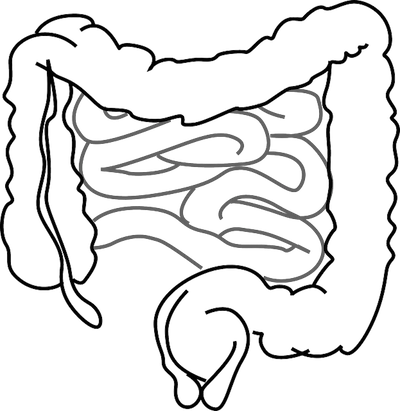
Inflammatory bowel disease (IBD) is a disease of the gut made up primarily of two diseases, primarily Crohn's disease and ulcerative colitis. In IBD, the gut and other parts of the digestive tract are attacked by the body's own immune system.  It is not clear how or why IBD occurs but there is a significant amount of research looking at this. It is known that the human gut microbiome goes through a lot of changes from birth through the first few years of life before stabilizing into the “adult” composition. Therefore, looking at the first years of life could be critical for understanding IBD. Researchers in Canada, in a study published by Clinical Gastroenterology and Hepatology, set out to determine if birth by Cesarean section increases likelihood of IBD.

The logic behind this hypothesis is that birth by vaginal delivery would expose the infant to the mother’s vaginal bacteria, which could possibly be essential in the development of the infant’s own microbiome. To study whether C-sections are a risk factor for IBD, the researchers gathered data from the University of Manitoba IBD Epidemiology Database, which keeps health records of all Manitobans diagnosed with IBD between 1984 and March 2010. These records were matched with birth and maternal health records. 1,671 IBD patients were linkable with mothers and therefore used for analysis. 10,488 matched controls were also used.

Analysis showed that IBD patients were no more likely to be born by C-section than the controls. Additionally, urban rather than rural residence was associated with higher instances of IBD. Within families, the likelihood of IBD was not different between C-section and non-C-section siblings.  

In conclusion, there does not appear to be an increased risk for inflammatory bowel disease if a child is born by C-section. Factors such as breastfeeding, socioeconomics, living environment, pets, etc. are still being analyzed for possible contribution to gut microbiome dysbiosis.

Please email blog@MicrobiomeInstitute.org for any comments, news, or ideas for new blog posts.

The views expressed in the blog are solely those of the author of the blog and not necessarily the American Microbiome Institute or any of our scientists, sponsors, donors, or affiliates.

Cigarette smoke changes the gut microbiome

When we talk about smoking cigarettes, we usually discuss the harmful effects that it has on our lungs, mouth, skin, and other parts of the body. However, we don't often talk about the gut even though cigarette smoke is the best-known environmental risk factor for Inflammatory bowel diseases (IBD), Crohn’s disease and ulcerative colitis.  While the exact mechanism for why people get these diseases is not yet known, it is recognized that a dysbiosis of the gut plays a contributing role to the onset of these conditions. A research team in Germany investigated the effects that cigarette smoke exposure had on the mucus layer and the microbes in the gut.

The scientists exposed mice to cigarette smoke or air for a period of 24 weeks. They found there was a shift in the microbial community in the caecum and distal colon after exposure to smoke. Specifically, there was an increase in Lachnospiraceae in the colon however it remained the same in the ileum, the last part of the small intestine.

They also found that smoke exposure led to changes in mucin exposure. Mucin is a type of protein that is known for producing gels that act to lubricate and protect parts of the body, both internal and external. The most common mucins are Muc2, Muc3, and Muc4. Muc2 for example is a protein that is secreted onto the mucosal surfaces of the large intestine and serves as a protective barrier for the epithelium. In this study, they found that Muc2, Muc3, and Muc4 gene expression was altered after cigarette smoke exposure.

The authors hypothesize that cigarette smoke affects the immune system in the ileum and may lead to the inflammation associated with Crohn’s disease. Overall, this study found that exposure to cigarette smoke had a profound effect on the gut bacteria and mucin composition in the mouse. While this was not done in humans, the same effects would likely be seen.

Please email blog@MicrobiomeInstitute.org for any comments, news, or ideas for new blog posts.

The views expressed in the blog are solely those of the author of the blog and not necessarily the American Microbiome Institute or any of our scientists, sponsors, donors, or affiliates.

Immune cells are educated in the gut to not attack beneficial bacteria

The gastrointestinal tract is made up of trillions of bacteria that are largely ignored by the body’s immune system.  Why is it that the body’s immune system knows to ignore these beneficial bacteria that are so important for our ability to live a healthy life? The answer to this question could play an important role in understanding how to maintain a healthy gut and how to treat diseases. Scientists led by Gregory Sonnenberg at Weill Cornell Medical College may have answered this question in a study published last week in Science.

The researchers studied T cells, cells that are made in the thymus and are trained there to kill-off foreign microbes and other intruders that make their way into the human body. But why don’t these T cells attack helpful bacteria in the GI tract? They found that the T cells are again educated in the gut to not attack beneficial bacteria but when this education is disrupted, it can lead to disease.  For example, inflammatory bowel diseases like Crohn’s disease and ulcerative colitis occur when the immune system attacks the GI tract and bacteria in the GI tract.

In the thymus, T cells that could attack the body are destroyed before they are released into circulation. In the gut, a type of cell called innate lymphoid cells (ILCs) educate the T cells to not attack beneficial bacteria. These ILCs had previously been found to make a physical barrier between the bacteria in the gut and the immune system.

In mice, they found that ILCs attacked T cells that were destroying beneficial bacteria and when they prevented this attack by ILCs on the T cells, severe intestinal inflammation resulted. They also looked at intestinal biopsies of young patients with Crohn’s disease. In the biopsies they found that the ILCs lacked specific molecules that are important for educating the T cells not to attack the bacteria in the gut. They found that a decrease in this molecule correlated with an increase in pro-inflammatory cells in children with Crohn’s disease.

The authors state that it may be possible to get rid of these T cells that are causing the inflammation and by doing so you may be able to help treat the disease.  By restoring this molecule (Major Histocompatibility Complex class II) that is preventing the education of the T cells, pro-inflammatory T cells may be reduced resulting in reduced intestinal inflammation.


Please email blog@MicrobiomeInstitute.org for any comments, news, or ideas for new blog posts.

The views expressed in the blog are solely those of the author of the blog and not necessarily the American Microbiome Institute or any of our scientists, sponsors, donors, or affiliates.

New info on fecal microbiota transplants for C. difficile and ulcerative colitis

"Fecal bacterial communities of recurrent [C. diff]  patients shift towards [healthy] fecal bacterial communities after FMT. Pre-FMT patient samples (red circle); post-FMT patient samples (green circles); trajectory of patient fe…

"Fecal bacterial communities of recurrent [C. diff]  patients shift towards [healthy] fecal bacterial communities after FMT. Pre-FMT patient samples (red circle); post-FMT patient samples (green circles); trajectory of patient fecal communities after FMT (blue line)."
Image and caption from the C. diff paper: Weingarden et al. Microbiome 2015 3:10   doi:10.1186/s40168-015-0070-0

Two important papers regarding fecal microbiota transplants (FMTs) were published last week.  The first was an examination of a patient’s microbiome over time after he or she undergoes an FMT to treat C. difficile.  The second showed the results of clinical trials that used FMTs in an attempt to treat ulcerative colitis.   The FMT papers, which are described below, improve our understanding of this procedure, which holds promise to treat various microbiome-based diseases.

The C. diff paper, published in the journal Microbiome, attempted to answer the question: Do the microbiome changes that occur after FMT remain long after the procedure?  We know that FMTs are highly effective in treating C. diff because they install a healthy microbiome that can crowd out the infection.  However, it is unknown if these new bugs that take hold are transient, or if they become permanent members of the gut.  The researchers sampled the microbiomes of FMT donors and recipient patients before and up to 84 days after an FMT procedure to treat C. diff.  They discovered that the recipients’ dysbiotic microbiomes stabilized quickly, and after just one day they closely resembled the donors’ microbiomes.  Continued measurements showed that the microbiomes deviated over the next few weeks, but that they remained healthy.

The colitis clinical trial, published in the journal Gastroenterology, attempted to discover if FMTs could treat ulcerative colitis.  Ulcerative colitis is widely considered to somehow be related to a dysbiosis in the microbiome, so can FMTs from healthy donors treat this disease?  The study was a double blind randomized clinical in which 48 people suffering from ulcerative colitis either received stool from healthy donors (treatment) or just an FMT of their own stool (control).  7/23 patients who received stool from a healthy donor were in remission after 12 weeks, while 5/25 patients who received their own stool were in remission at that time.  Unfortunately, this is not a clinically significant result based on the number of patients involved.  The researchers measured the bacterial abundance in all of the patients microbiomes before and after treatment.  Before treatment the microbiomes all had some baseline similarity.  After treatment, though, the patients who responded to treatment from a healthy donor all had an increase in certain Clostridia, and the patients who responded to treatment from their own stool all had in increase in certain Bacilli, Proteobacteria and Bacteriodetes.  The researchers feel that this information warrants further study.

FMTs are an exciting new therapy that may be important in treating some really nasty diseases.  We do want to remind people, though, that it is still an unproven technique that should only be performed under the guidance of a doctor.  As we have written about before, the promise of the microbiome is what makes FMTs both attractive, but potentially dangerous at the same time.

Please email blog@MicrobiomeInstitute.org for any comments, news, or ideas for new blog posts.

The views expressed in the blog are solely those of the author of the blog and not necessarily the American Microbiome Institute or any of our scientists, sponsors, donors, or affiliates.

Understanding spatial relations of gut bacteria in ulcerative colitis patients

To sample the microbial communities in the gut, fecal samples are generally collected from an individual and DNA is sequenced to identify bacteria that are present. This is an overall effective method, however, it does not provide information of the specific spatial location of bacteria within the gut. In a study published in the journal Gut, researchers in Ireland looked to determine differences in the bacterial composition of specific regions of the large intestine between patients with ulcerative colitis and control patients.

Four volunteers undergoing routine colonoscopies were recruited to serve as the controls for this study. Five patients with ulcerative colitis (UC), who were undergoing colectomies, or surgical removal of the colon, were also involved in the study. Samples were taken at four locations in the colon in all individuals: the caecum, traverse colon, descending colon, and rectum. The four locations were sampled three times at three different levels: luminal brush, whole mucosal biopsy, and laser captured sample of mucus gel layer. A total of twelve samples were taken per individual.

After analysis of the many samples it was discovered that there was more variability between the bacterial compositions between subjects than there was within the different locations of an individual’s colon. The findings showed a difference between the luminal and mucus gel microbiota in both the controls and the ulcerative colitis subjects. Three bacterial families were common between this difference shared by controls and UC subjects, namely Bifidobacteriaceae, Peptostreptococcaceae, and Enterobacteriaceae being more abundant in UC patients.

This study has its limitations because of the small sample size, however the researchers state that the small sample allowed for extensive analysis of the individual samples. So what do the findings of this study mean for patients with ulcerative colitis? Better understanding of differences in the spatial relations of bacteria could lead to the modulation of microbial communities to help treat ulcerative colitis. 

Please email blog@MicrobiomeInstitute.org for any comments, news, or ideas for new blog posts.

The views expressed in the blog are solely those of the author of the blog and not necessarily the American Microbiome Institute or any of our scientists, sponsors, donors, or affiliates.

Viruses in the gut connected to inflammatory bowel disease

Drawing of a bacteriophage

Drawing of a bacteriophage

A new study has shown that the composition of viruses in the gut may play an important role in inflammatory bowel diseases (IBD).  If you’ve been reading the blog for a while, you’ve seen us write about something called the virome. The virome is the collection of viruses in the body and similarly to the microbiome, it may have profound affects on human health. This study led by scientists at Washington University in St. Louis and published in Cell is the first to correlate a disease with changes in a person’s virome.

IBD, specifically Crohn’s disease and ulcerative colitis (UC), are diseases that have been characterized by decreased bacterial diversity in the gut.  However in this study, the scientists found that patients with Crohn’s and UC showed greater diversity of viruses than healthy individuals.  This suggested that viruses played a role in the disease.

The team of scientists studied individuals in Boston, Chicago, and the United Kingdom with the disease. They took stool samples from patients with UC and Crohn’s and sequenced their viral DNA. They compared this to the viruses in stool samples from healthy individuals living in the same areas and households. Patients with the disease had a higher number of viruses than those without IBD. Specifically, they found that Crohn’s and UC patients had higher levels of Caudovirales bacteriophages (viruses that infect bacteria) that were specific to each disease.

Further research is needed to better understand the relationship between the virome and the microbiome but as we see from the increase in bacteriophages, there is certainly a relationship between these two systems. While the authors state that it does not look as if changes in the virome were secondary to changes in bacterial populations, it is not yet clear if changes in the virome are the result of bacterial alterations in the gut or if it may lead to microbiome changes - or a combination of the two.  This study is the first of its kind to show a connection between disease and the virome and I think we are going to see several more studies in the coming years showing this type of correlation with disease.  While we generally think of viruses as causing infections like influenza, their impact on chronic disease may be vast.  

Please email blog@MicrobiomeInstitute.org for any comments, news, or ideas for new blog posts.

The views expressed in the blog are solely those of the author of the blog and not necessarily the American Microbiome Institute or any of our scientists, sponsors, donors, or affiliates.