Corynebacterium

Operating room bacteria colonize infants’ guts after C-sections

operation-theatre-555088_640.jpg

Newborns are a great study subject in the field of microbiology, because scientists are still discovering how the microbiome develops and what factors affect it. In human infants, it has been proven that vaginal birth exposes infants to bacteria that are different from those received by the mother through C-section. Babies born by vaginal delivery have gut bacteria correlated with vaginal bacteria, while babies born by C-section have gut bacteria correlated with human skin bacteria. For babies born by C-section, the sources of the human skin microbes that are acquired are still unknown.  In a study published by Microbiome, a group of scientists tested the hypothesis that the operating room environment contains human skin bacteria that could be seeding the gut microbiome of C-section born babies.

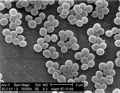
To test their hypothesis, the researchers collected samples from 11 sites in four operating rooms from three hospitals in New York City, NY and San Juan, PR. Of the 44 operating room samples that were collected, 68% of the samples contained a sufficient number of bacterial DNA samples for sequence analysis. After analyzing the bacteria collected, it was found that all samples contained human skin bacteria, with Staphylococcus and Corynebacterium being the greatest in quantity. Lamps on the operating bed and baby crib showed higher abundances of these bacteria relative to the other sampling sites. The scientists confirmed that the samples collected were more similar to human skin microbiota than other body sites, by comparing the samples to oral, fecal, and vaginal database samples.       

Even though operating rooms are supposed to be spotlessly clean and germ-free, this study shows that there are still dust particles containing human skin, and therefore human skin microbiota, samples. These samples could be from people moving in and out of the operating room during a C-section, or it could come from the people cleaning the OR. Either way, the human skin bacteria in the operating room most-likely are what influences the infant’s developing gut microbiome. 

Please email blog@MicrobiomeInstitute.org for any comments, news, or ideas for new blog posts.

The views expressed in the blog are solely those of the author of the blog and not necessarily the American Microbiome Institute or any of our scientists, sponsors, donors, or affiliates.

We all emit our own 'microbial cloud'

Every individual has a microbiome compiled of millions of bacteria, fungi, viruses, and other microorganisms that is unique for each one of us. Whenever we travel to a new location and sit down or touch something, we are spreading our microbiome to that new location. A lot of research has gone into this phenomenon and is called the microbiome of the built environment. A new study out of the University of Oregon has expanded on this understanding and has described what they call a “microbial cloud”.

The scientists found that individuals not only spread their microbiome to new locations through direct contact but the microorganisms on our body are also dispersed into the air making up this microbial cloud. To better understand this, the scientists had 11 individuals sit in an enclosed room for 4 hours and they analyzed the DNA from the bacteria in the air. They found that when each individual sat in the room, there were thousands of bacteria in the room and everyone’s was distinct. They were able to identify specific characteristics of the people such as if it was a man or a woman.

The bacterial combinations found in the room could be linked back to specific individuals even after the person inhabited the room for only 4 hours. There were specific groups of bacteria like Streptococcus, often found in the mouth, as well as Propionibacterium and Corynebacterium, often found on the skin, that were most useful in identifying the individuals. While these bacteria were found around all the study participants it was the combination of bacteria that was key to identifying the individuals.

This finding could have several important applications. One often-discussed application of the microbiome is its use in forensic applications. It may be possible to use this ability to identify people and know if they were in a room or not to see if someone committed a crime, though it is not clear if it will be possible to identify people in a crowd of other individuals. Other applications include understanding the spread of infectious disease between individuals and within buildings. This is an exciting new development and I am certain we will see more research looking at our microbial clouds in the future.

Please email blog@MicrobiomeInstitute.org for any comments, news, or ideas for new blog posts.

The views expressed in the blog are solely those of the author of the blog and not necessarily the American Microbiome Institute or any of our scientists, sponsors, donors, or affiliates.

Study suggests penile microbiome can transmit bacterial vaginosis by sexual intercourse

Bacterial vaginosis (BV) is a microbiome-based disease characterized by a lack of Lactobacillus in the vagina.  We have covered this disease with multiple blog posts and encourage any interested readers to search for these blogs to learn more.  One outstanding question regarding BV is how sexual intercourse affects the disease.  One prevailing thought is that the penis can actually be colonized by BV-associated bacteria, and that through sexual intercourse it can be spread between partners.  A new paper published last week in mBio suggests this is true.

The researchers measured the penile microbiomes of 165 uncircumcised, black men from Uganda, as well as diagnosing BV status in their female partners.  The BV status was measured by Nugent score, which is a bacterial staining technique that basically measures the amount of anaerobic bacteria in the vagina (non-Lactobacilli).  The stain produces a score between 1-7 with 1 being healthiest and 7 being least healthy (mostly anaerobic bacteria).  After measuring the penile microbiomes, the scientists were able to be categorize them into 7 different community state types (CST1-7).  These community state types varied from 1 to 7 in terms of both overall abundance and composition, with CST1 having the lowest density of bacteria and the lowest diversity while CST7 had the highest density and the highest diversity of bacteria.

The scientists compared the female partner’s BV status with the men’s community state type, and noted that having a CST1-7 on the penile microbiome corresponded with a higher likelihood of the female partner being diagnosed with BV.  Two genera of bacteria, Corynebacterium and Staphylococcus, on the penile microbiome were associated with healthy vaginal flora, whereas Dialister, Mobiluncus, Prevotella, and Porphyromonas were associated with BV.  Interestingly penises that included Lactobacillus and Gardnerella, genera associated with healthy vaginas and BV vaginas, respectively, were not statistically associated with BV status.  Overall, men with CST4-7 were significantly more likely to have a sexual partner with BV, and had more BV associated bacteria colonizing their penises.  In addition, men with more than one sexual partner were more likely to have CST4-7, and again, their partners more likely to have BV.

It appears that men’s penises, especially uncircumcised ones, can be vectors for bacterial transmission.  This simple fact should make us reconsider BV as an STD, and actually fits in well with another that has shown promiscuity is a risk for BV.  It is likely that circumcision and condom would decrease BV transmission rate, as they do other STDs, but until a paper comes out that studies this connection no one can say for sure.

Please email blog@MicrobiomeInstitute.org for any comments, news, or ideas for new blog posts.

The views expressed in the blog are solely those of the author of the blog and not necessarily the American Microbiome Institute or any of our scientists, sponsors, donors, or affiliates.

Understanding the nasal microbiome

Electron micrography of Staphylococcus aureus.

Electron micrography of Staphylococcus aureus.

The nasal microbiome remains largely unstudied despite its potential importance to many diseases, such as rhinosinusitis, allergies, and staph infection (incuding MRSA).  Staphylococcus aureus is probably the most well-known nasal resident, but simple questions, such as which species of bacteria are most prevalent in the nose, are still not answered.  Understanding all the residents of the nasal microbiome, the influence of our genetics and the environment on defining their populations, and the influence each one has on others may be critically important to preventing diseases such as staph infection, and more research is needed.  Fortunately, a new study out of Johns Hopkins that investigated sets of twins shed light on many of these questions, and was published in Science Advances last week.

The scientists sequenced the nasal microbiomes of 46 identical and 43 fraternal pairs of twins.  First, thy learned that these people’s nasal microbiomes could be classified into 7 different phenotypes or community state types (CST) which broadly described their nasal microbiomes.  These 7 types are defined by their most abundant bacteria, and are as follows: CST1 – S. aureus, CST2 - Escherichia spp., Proteus spp., and Klebsiella spp., CST3 - Staphylococcus epidermidis, CST4 - Propionibacterium spp., CST5 - Corynebacterium spp., CST6 - Moraxella spp., and CST7 - Dolosigranulum spp.   The most common CTS was CTS4 with 29% of the sampled population having that CTS, whereas CTS4 was the least popular, coming in at 6% of the individuals tested.  The researchers noted that many of these bacteria, such as Proteus, were not considered to be important to the nasal microbiome at all, so their dominance in some noses was surprising.  The scientists learned that genetics plays nearly no role in the microbiome community composition, but does influence the overall microbiome population.  In addition, gender influenced the overall population, with women having about half as many total bacteria in their noses as men.

With regards to S. aureus, while it existed in 56% of the individuals studied, it was associated with other bacterial.   For example, the researchers discovered that Dolosigranulum, and Propionibacterium granulosum were negatively correlated to the existence of S. aureus, whereas S. epidermidis was positively correlated with S. aureus abundance.  This lends itself to the idea that specific bacteria can create colonization resistance against S. aureus, and thus could be used to prevent the disease.  The researchers suggest a probiotic should be tested for its therapeutic value in preventing S. aureus colonization, and hopefully they move forward with those trials.

Please email blog@MicrobiomeInstitute.org for any comments, news, or ideas for new blog posts.

The views expressed in the blog are solely those of the author of the blog and not necessarily the American Microbiome Institute or any of our scientists, sponsors, donors, or affiliates.

The nasal microbiome of infants may impact risk of developing asthma

Many lower respiratory illnesses have been shown to associate with specific lung, throat and nasal bacteria, but the role of the microbiome is still unclear, and mechanisms for the connection have yet to be proven.  Of particular interest is asthma, which affects around 7% of people in the US, and increases a person’s risk for many other conditions.  While it is normally diagnosed in toddlers, scientists believe that the groundwork for the disease is actually laid during infancy.  With that in mind, researchers in Australia performed the first longitudinal study of infants’ nasopharyngeal (nose and throat) during the first year of their lives, and tracked episodes of respiratory illness during that time.  They discovered a strong connection between the microbiome and respiratory illness, including asthma, and last month published their results in Cell Host and Microbe.

The researchers collected nasopharyngeal microbiome samples from 234 infants at different time points during their first year of life.  Most infants’ microbiomes were dominated by the following species: Moraxella catarrhalis, Streptococcus pneumoniae, Staphylococcus aureus, Haemophilus influenzae, and Alloiococcus otitidis.  Interestingly, this was true for infants regardless of birth delivery mode (i.e. cesarean or vaginal) as well as length of breast feeding.  In contrast, having a furry animal in the house tended to increase the abundance of Streptococcus, but having older siblings tended to decrease it.  In addition, there were strong seasonal effects on the microbiome, with Haemophilus being associated with the summer, and Moraxella the winter.  In children with respiratory illness, Haemophilus, Moraxella, and Streptococcus were most frequently measured, and Staphylococcus, Alloiococcus, and Corynebacterium least frequently measured.

When the scientists compared their results with the asthma outcomes of the children at 5 years old they noticed one significant trend.  Colonization by Streptococcus at around 2 months old, which was asymptomatic at the time and occurred in 14% of infants tested, was strongly connected to chronic wheezing (itself an indication of asthma) at 5 years old.  They suggest that the developing airways in infants may be especially vulnerable to Streptococcus.

This long term study does a really nice job of defining how the microbiome grows and develops in the airways of infants – something which previously hadn’t been performed at such a large scale.  While this study alone does not figure out exactly what the microbiome’s role is in childhood respiratory illnesses, it does provide a baseline for future studies to work off of.   

Please email blog@MicrobiomeInstitute.org for any comments, news, or ideas for new blog posts.

The views expressed in the blog are solely those of the author of the blog and not necessarily the American Microbiome Institute or any of our scientists, sponsors, donors, or affiliates.

New study suggests S. aureus and skin dysbioses cause eczema

Atopic dermatitis, also known as eczema, is a skin inflammation and rash that has an enigmatic cause.  There are many genetic and environmental risk factors involved, but to date the exact triggers and mechanisms that cause this autoimmune response are unknown.  Importantly, a growing percentage of infants and toddlers are developing this disease, which itself is a known risk factor for other autoimmune diseases like asthma and allergies.  Discovering the cause of atopic dermatitis is an important endeavor, because it may lead to a cure for a number of other diseases.

Staphylococcus aureus has long been associated with atopic dermatitis.  It appears to occur at relatively high abundances in the areas of skin that are affected.  Then again, S. aureus is a one of the most common skin microbiome bacteria (it is ubiquitous around the world), and it has yet to be definitively connected to the disease.  In addition, mouse models for many skin diseases, including this one, do not exist or are insufficient, so controllably studying the atopic dermatitis is difficult.  Recently though, a team of scientists from Japan and the NIH developed a mouse model for atopic dermatitis, and made a new discovery that showed S. aureus can indeed drive skin inflammation.  They published their results in Cell immunity.

The scientists were studying how a specific genetic mutation in mice affected bones and hair follicles when they serendipitously realized that it was causing eczema in the mice after around three weeks.  When they investigated the skin microbiomes of these mice, as well as normal mice, they realized that right around the time that the eczema was appearing in the mice, these mice’s skin microbiomes drastically shifted.  First, a bacterium called Corynebacterium mastitidis emerged, followed by S. aureus a few weeks later, which was coincident with the presentation of the worst symptoms.  Of note, species of Corynebacteria are associated with eczema in humans, much like S. aureus.

Next, the researchers then performed a series of experiments by providing mice with antibiotics in an effort to combat the dysbiosis.  When newborn mice with the genetic modification were treated with antibiotics they never developed eczema at all.  Moreover, genetically modified mice that were in the midst of the rash that were treated with antibiotics had their eczema subside soon after.  In addition, genetically modified mice that were taken off of antibiotics had eczema emerge shortly thereafter.  Strikingly, in all of the above situations changes in the skin microbiome corresponded with the disease state: a lack of S. aureus and high diversity were associated with healthy skin, and the emergence of S. aureus and a lack of diversity were associated with the disease.  Finally, when S. aureus was inoculated onto the skin of genetically modified mice, they developed eczema rapidly.

The researchers performed a number of other experiments to try and tease out the mechanism by which S. aureus causes atopic dermatitis.  Their results show that it appears a combination of genetic and environmental factors that affect the skin may be important in defining an individual’s risk for the disease.  Regardless, it appears that S. aureus is a major culprit in causing eczema, so future therapies that eradicate the bacteria, or at least decrease its abundance should be considered.

Please email blog@MicrobiomeInstitute.org for any comments, news, or ideas for new blog posts.

The views expressed in the blog are solely those of the author of the blog and not necessarily the American Microbiome Institute or any of our scientists, sponsors, donors, or affiliates.