Helminths may provide therapeutic benefit to treat brain disorder

We’ve recently talked about a few articles that have studied helminth infection with respect to the microbiome, and how these infections could possibly confer some therapeutic benefits.  Another recent study conducted by researchers at Duke University reinforces these findings.  Autoimmune and inflammatory disorders appear to be more common in developed societies, and many have suggested that the microbiome is a major driver of these changes to our immunity.  These investigators wanted to assess whether or not helminths – which have a lot of influence on the immune system – had any effect in modulating the brain immune system in the context of living conditions and early-life infection, as this has been shown to result in neurodevelopment disorders. 

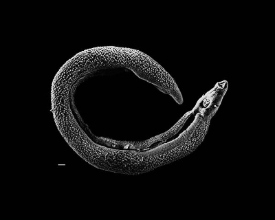
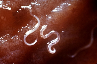
In this study, male and female rats were infected with a H. diminuta cystercircoid rat tapeworm a few weeks prior to breeding.  The rats were segregated by living conditions, housed in either dirty colonies (or “farm-like” environments), where no water or air filtration was provided) or standard clean pathogen-free laboratory conditions.  The offspring in both environments were delivered helminths, and the males were infected with E. coli early in life. 

Later in adulthood, the immune systems of the progeny animals were challenged by lipopolysaccharide (LPS) inductions in learning tests, and brains were collected shortly after to examine changes in molecular immune responses.  Exaggerated immune responses were observed in rats that were infected with E.coli early in life in the standard clean lab conditions.  Alternatively, the cohort that lived in the farm-like conditions did not experience an increase.  Both groups were infected with helminths.

To narrow down further, the researchers examined the impact of helminths alone in rats housed under clean pathogen-free laboratory conditions.  Indeed, cytokine responses in rats infected with E.coli were reduced in the animals whose mothers were infected with helminths before giving birth.  In addition to immunologic modulation, helminth infections in adult rats where shown to reduce memory deficits that are common following E. coli infection, suggesting helminth infection played a role in modulating developmental disorders due to bacterial infection. 

The helminths also had an effect on the microbiomes of the rodents.  16s rRNA sequencing revealed an average 25% shift in microbiome composition of animals infected with helminths (with a predominant shift of Bacilli to Clostridia).  Rats that were infected with E. coli early in life experienced a microbiome composition shift in adulthood, as more harmful Bacteroidetes species were found in adults.  Interestingly, this observation was not found in those who were E.coli infected but also infected with helminths, suggesting helminths prevented this composition shift. 

Overall, these findings suggest that helminths could provide therapeutic benefit, especially after infection early in life.  It will be interesting to see how this research can translate to human models, especially by narrowing down bacterial infections that could harm or benefit development.  Understanding what drives these developmental complications could have immense health benefits for the public. 

Please email blog@MicrobiomeInstitute.org for any comments, news, or ideas for new blog posts.

The views expressed in the blog are solely those of the author of the blog and not necessarily the American Microbiome Institute or any of our scientists, sponsors, donors, or affiliates.

Fecal microbiota transplant as a treatment for MRSA enterocolitis

Fecal microbiota transplants (FMTs) are most commonly used for treating Clostridium difficile infection, an often lethal bacterial infection of the gut. However, there have been many hypotheses that FMTs could be used to treat other conditions that result in a dysbiosis of the microbiota. A new study published in BMC Infectious Diseases suggests that FMTs could be used to treat enterocolitis, infection of the gut, that is a result of Methicillin-resistant Staphylococcus aureus (MRSA).

The most common treatment for this to date has been antibiotic treatment, specifically vanomycin, but the results of how this impacted the microbiota were not measured. In this new study, 5 patients with enterocolitis as a result of MRSA were given FMTs, the infusion of fecal preparation into the GI tract of the patient from a healthy donor. After administration of the FMT, all 5 patients were cured of the MRSA enterocolitis showing no symptoms. MRSA in the feces was also eliminated after FMT.

They also measured the microbiome of patients undergoing the treatment. They found that prior to treatment, patients with MRSA enterocolitis had decreased numbers of species in the gut and S. aureus reached almost half of all intestinal flora.  After the FMT, the microbiome of the recipient trended closer to the microbiome of the donor and alleviated symptoms. 

While there remain concerns with the use of FMTs, there are certain instances where there are few options for treatment and the administration of a new microbiome from a donor fecal sample remain the most promising. While this was only a study of 5 patients at one hospital in Singapore, the investigators suggest FMT as a first-line measure treatment for enterocolitis resulting from MRSA. 

Please email blog@MicrobiomeInstitute.org for any comments, news, or ideas for new blog posts.

The views expressed in the blog are solely those of the author of the blog and not necessarily the American Microbiome Institute or any of our scientists, sponsors, donors, or affiliates.

Burn injuries lead to changes in the gut microbiome

One of the leading causes of death among burn injury patients is infection, and specifically sepsis. It is believed that in individuals with compromised immune systems, sepsis is caused by a bacterial infection that leads to an inflammatory response throughout the whole body. The powerful inflammatory response changes blood pressure and causes cell stress in the body, which is most often fatal. Additionally, many studies suggest that Gram-negative (this classification is based on cell membrane composition) bacterial infections have something to do with sepsis. Published in Plos One, researchers in Chicago studied burn patients and mice to see if burn injury changed the gut in such a way that it becomes easier for Gram-negative bacteria to thrive.

Fecal samples were collected from 4 burn patients at Loyola University Medical Center. The total body surface area (TBSA) of burns for each patient was 25%, 32%, 44%, and 57%. A control group of 8 patients was used as well, made up of burn patients whose TBSA was less than 10%. A similar mice experiment was set up as well.

After comparing the fecal samples of the significantly burned patients and the controls, they found that fecal microbial richness was significantly higher in control patients. The burn patients with the highest TBSA had the most similar communities, and after 11 days the community of the 32% patient changed to more closely resemble that of the 44% and 57% patients. Sadly, the burn patients with 32%, 44% and 57% TBSA died from sepsis. Upon examination of the microbes collected in fecal samples from the burn patients, however, a relative abundance of bacteria was found from the family Enterobacteriaceae, which are Gram-negative.

The effects of burn injury were studied in mice as well, in order to determine if the human effects were reproducible in mice, if similar microbes are found in the mice gut, and if different locations in the gastrointestinal tract had different community characteristics. The small and large intestines were found to have very different microbiome composition. The abundance of Enterobacteriaceae was higher in the small intestine than the low intestine, but the abundance decreased from day one to day three. 

The intestinal wall can be injured because of burns, and if the ability for things to move through it is easier, then harmful bacteria may be able to move into the circulatory system and increase the risk of an inflammatory response. The proteins that hold cells together in the gut were studied, and they were found to be decreased in burn injury mice. This occurred at the same time as the increase of Enterobacteriaceae. While an established connection cannot be made yet, this study will hopefully lead to further research of the connection between Gram-negative bacteria and sepsis. 

Please email blog@MicrobiomeInstitute.org for any comments, news, or ideas for new blog posts.

The views expressed in the blog are solely those of the author of the blog and not necessarily the American Microbiome Institute or any of our scientists, sponsors, donors, or affiliates.

Randomized clinical trial shows probiotic may not be an effective treatment for colic

Many families have experienced colicky infants who have excessive and inconsolable crying.  The cause of this behavior is largely unknown, however it is beginning to be linked to a variety of diseases, including allergies and gastrointestinal disorders.  Many remedies have been suggested to help assuage these infants, including probiotic therapies, but thus far the evidence of their efficacy is unknown.  Researchers in Finland put one of the probiotic therapies, using Lactobacillus rhamnosus GG (LGG), to the test and conducted a double blind randomized clinical trial to discover whether it decreased colic.  They published their results last week in Nature Pediatric Research

The scientists studied 30 colicky infants in the study, who were split evenly into a probiotic group and a control group.  The mothers of the probiotic group orally administered LGG to their children once a day for 28 days, while the mothers of the control group orally administered a placebo.  During this time the mothers kept diaries of how long the child cried, as well as collected stool samples for microbiome testing.  The results showed that the probiotics did not alter the amount of crying for each infant when compared to the placebo group.  In addition there was no statistical difference in the microbiome’s of both groups.

Unfortunately for the families of colicky infants, this study did not show that LGG was an effective colic therapy.  There are other studies that conflict with this one though, so perhaps different types of bacteria, or larger doses could improve efficacy.  The relationship between the microbiome and colic is unclear, however, given the recent advances in gut-brain axis research, we would not be surprised if the two are connected.


Please email blog@MicrobiomeInstitute.org for any comments, news, or ideas for new blog posts.

The views expressed in the blog are solely those of the author of the blog and not necessarily the American Microbiome Institute or any of our scientists, sponsors, donors, or affiliates.

Yogurt may help combat colitis

Editor's note:  The following work comes out of Wendy Garrett's lab at Harvard.  Wendy will be an upcoming guest on the podcast.  If you have questions for her, feel free to email or call, and we will ask her on the show. 

A key signature of colitis is the build-up of reactive oxygen species (ROS) in the colon due to chronic inflammation.  One of major functions of ROS is actually to act as an antibiotic, and destroy any foreign bacteria that may exist at the site of inflammation.  However, ROS are known to be toxic to the host as well, and their high concentrations in colitis are likely a major contributor to the disease.  With this in mind, researchers at Harvard studied how probiotics from yogurt could ameliorate the disease by disposing the excess ROS.  They published their results last month in the Proceedings of the National Academy of Sciences.

The researchers had previously determined that a common yogurt that contained 5 strains of bacteria was helpful in decreasing symptoms in multiple mouse models of colitis.  In this study though, they identified Lactococcus lactis as being the most important of these strains in treating colitis symptoms.  They then compared the genome of L. lactis with the other strains in the yogurt and determined that a specific gene that codes for the enzyme, superoxide dismutase (SOD), which is capable of breaking down superoxide, an ROS, may be imparting L. lactis’ beneficial effects.  In order to support this hypothesis, the scientists showed that when this gene was removed from L. lactis the bacteria no longer reduced colitis.  They took this notion a step further and showed that superoxide levels were in fact decreased in vitro when combined with lysed L. lactis.  Finally, the scientists showed that the L. lactis must actually be lysed in the colons in order to release its SOD, destroy superoxide, and reduce colitis: when the scientists attempted to deliver SOD on its own to mice with colitis it was not as effective, and caused diarrhea.

This study is really interesting for two reasons.  The first is that it shows yogurt, like Activia, may be very helpful in dealing with colitis.  The second reason though, is that it shows a new system for deliverying SOD to a site of inflammation: via bacteria.  As they showed in the paper, simply using SOD was not effective, but using the bacteria as a vehicle for SOD, and then lysing it at the site was an effective means of drug delivery.  This has many important implications because ROS are important contributors to a variety of diseases.  

Please email blog@MicrobiomeInstitute.org for any comments, news, or ideas for new blog posts.

The views expressed in the blog are solely those of the author of the blog and not necessarily the American Microbiome Institute or any of our scientists, sponsors, donors, or affiliates.

Microbiome composition of children infected with helminths

Schistosoma parasite worm, otherwise known as a blood fluke.

Schistosoma parasite worm, otherwise known as a blood fluke.

While much has been explored regarding the microbiome’s role in nutrition and immunology, more research is needed in uncovering interactions between the host microbiome and infection as this represents a high unmet medical need.  A few weeks ago we talked about a study that describes an interesting relationship between helminth infection and sensitivity to insulin in Indonesia.  Helminth infections are also prevalent in sub-Saharan Africa, where many children become exposed to Schistosoma haematobium, causing schistosomes and dramatically affecting childhood health and development.  Researchers from Africa sought to investigate whether there were significant differences in microbiome composition between children who were infected with S. haematobium and those who were not.  Furthermore, investigators explored whether praziquantel (PZQ) – an effective agent that kills schistosome worms – has any influence on the human microbiome composition of infected patients. 

Stool samples were collected from 139 pediatric patients from six months to 13 years old, and groups were segregated following proper diagnosis of S. haematobium infection.  DNA was extracted from the samples and microbiota were characterized using 16S rRNA sequencing.  Overall microbiota compositions were similar across sex and all age groups, and Bacteroidetes phyla were found to be most abundant.  However, there was a significant difference in operational taxonomic unit (OUT) microbiota clusters (a measurement that categorizes bacteria colonies) between infected and non-infected groups.

In the next experiment, researchers investigated the microbiomes in the 62 patients who took the PZQ therapy, comparing microbiota after 12 weeks of treatment to their baseline compositions (i.e., prior to PZQ administration).  Interestingly, there was no statistical difference in microbiota composition in patients between pre and post administration time points. 

Though this study did not necessarily present breakthrough findings, the researchers presented more data that will assist doctors and clinicians to better understand helminth infection with respect to the microbiome.  Learning more about the exact differences in microbiota composition between infected and non-infected children will advance our understanding of interactions that could potentially lead to a novel and much-needed therapy.  

Please email blog@MicrobiomeInstitute.org for any comments, news, or ideas for new blog posts.

The views expressed in the blog are solely those of the author of the blog and not necessarily the American Microbiome Institute or any of our scientists, sponsors, donors, or affiliates.