Common yeast may trigger celiac disease onset

Candida albicans growing in petri dish

Candida albicans growing in petri dish

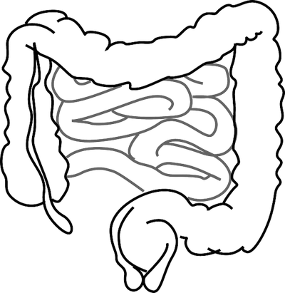
Celiac disease is a serious autoimmune disorder in which gluten, found in wheat, rye, and barley, triggers an immune response that damages the absorption capabilities of the intestines. The AMI has covered this topic in a few previous blogs (click celiac disease tag below) in relation to autoimmune disorders and possible bacterial triggers. One contributing factor to the celiac disease response is due to the protein gliadin, which is found in gluten. Gliadin, along with transglutaminase (which is a human protein that binds to, and deaminates gliadin), trigger a T Cell response that leads to the inflammation and tissue damage.

The yeast Candida albicans is a common gut commensal that is linked to inflammatory bowel diseases and vaginal infections.  This yeast also binds with transglutaminase, using a protein called Hwp1, in an identical fashion as gliadin.  This results in the bacteria’s strong binding to the intestinal wall, where it triggers an autoimmune response to destroy the yeast.  

Researchers in France hypothesized that the similarity between gliadin's and C. albicans' binding to transglutaminase may result in a similarity in the body's response to these two things.  In essence, they suggested that gluten ‘tricks’ the body into an immune response because it 'looks' similar to C. albicans.  

In the study, recently published by Plos One, blood cultures from 87 adult patients with celiac disease and 41 patients with C. albicans infection were collected.  The scientists then isolated the body's natural antibody for Hwp1 and measured its response to both gliadin and Hwp1.  They discovered that gliadin also binds to Hwp1's antibody, meaning that it should elicit the same immune response as Hwp1.  Therefore, the body should mount an immune response for gluten that is characteristic of C. albicans infection, and this response could manifest itself as celiac disease.

The significance of this study is that it comes closer to finding a cause and prevention of celiac disease. The T cell immune response that results from transglutaminase binding to gliadin could initially be triggered by a C. albicans yeast infection. This may explain why some people only become gluten sensitive later in life - perhaps it only occurs after they have a C. albicans infection and the body builds up antibodies for this yeast. This is another example of how microbes found in healthy individuals can be harmful when homeostasis is not controlled. 

Please email blog@MicrobiomeInstitute.org for any comments, news, or ideas for new blog posts.

The views expressed in the blog are solely those of the author of the blog and not necessarily the American Microbiome Institute or any of our scientists, sponsors, donors, or affiliates.

Antibiotic use in livestock is increasing and leading to greater antibiotic resistance

We’ve talked extensively about antibiotic resistance on the blog but we haven’t focused much on the impact that antibiotics given to livestock have on humans. Farmers give low doses of antibiotics to farm animals in order to not only prevent illnesses in their animals, but also to promote growth within their livestock. Animals being produced for food account for about 80% of antimicrobial use in the United States and bacteria in the animals become resistant to these antibiotics in the same fashion that they do in humans. As antibiotic use rises, more bacteria are becoming resistant and these bacteria are passed from animals to humans through the environment, consumption, and direct contact. An international research team from Europe, India, Africa, and the United States mapped the global consumption of antimicrobials in livestock.

The results were published in the Proceedings of the National Academies of Science (PNAS) and compared the use of antibiotics in livestock in 2010 to the projected use in 2030. The authors found that the global use of antibiotics in livestock will increase 67% in the 20 years between 2010 and 2030. This is largely a result of an increase in demand for meat by middle class individuals in countries like Brazil, Russia, India, China, and South Africa (BRICS). These BRICS countries have shifted their livestock production systems to be more cost-effective by increasing the use of antimicrobials to ensure the health of their animals and to promote growth.

This increase in antimicrobial use in animals is a compounded annual growth rate of 2.6% annual which is almost three times the annual growth rate of the human population (.98%) during the same time period. The authors found that in 2030, if new regulations are not put into place, approximately 30% of all antimicrobial use will be accounted for in the livestock industry in China.

Many countries, and specifically those in the developing world, do not regulate the use of antimicrobials in livestock production. While directly linking antibiotic use in animals to drug resistant infections in humans is very complex, it can be inferred that increase in antibiotic use leads to antibiotic resistant bacteria, and we have seen evidence of this in practice. In countries like India, where bacterial diseases are very prevalent and a major public health concern, antibiotics are a key factor in fighting these illnesses.  Increased resistance to bacteria by increased use of antibiotics in farm animals will only increasingly prevent the effectiveness of antibiotics in humans.  

The study authors call for global action to decrease the use of antibiotics used in animals that are raised for meat consumption. While the authors do state that this analysis was based on limited available data, largely in developed nations, the global trends of antibiotic use in livestock is concerning. 

Please email blog@MicrobiomeInstitute.org for any comments, news, or ideas for new blog posts.

The views expressed in the blog are solely those of the author of the blog and not necessarily the American Microbiome Institute or any of our scientists, sponsors, donors, or affiliates.

Breastfeeding associated with intelligence later in life

 Positive association between breastfeeding and intelligence, between three tiers of family income.  Five IQ points is 1/3 of a standard deviation. 


Positive association between breastfeeding and intelligence, between three tiers of family income.  Five IQ points is 1/3 of a standard deviation. 

Editor’s note: I would like to tread very lightly on this topic because of the complexity of the relationships between all the factors discussed and the implications a study like this has.  I probably would not have written about it at all, had it not been published in such a prestigious medical journal, and had been as comprehensive as it was.

Breast milk is the ultimate pre- and probiotic.  It is essential in developing infants’ microbiomes by inoculating and enriching their guts in certain bacterial species.  There have been a number of studies showing alterations of the gut microbiomes of infants that are formula fed, and other studies showing that formula feeding results in a higher risk of asthma and allergies later in life.  The complicated relationship between breastfeeding, the microbiome, and phenotypes like autoimmune diseases are not understood at a mechanistic level.  Still though, it appears that formula feeding, rather than breastfeeding, may have long term consequences for the health of a child.

To that end, there had been a few small scale studies that demonstrated a general association between breastfeeding an IQ.  Just last week, a new study, much more comprehensive than any previous one, delved into this topic and found the same result: duration of breastfeeding was positively associated with IQ, educational level, and income.  The results of this study were published in The Lancet.

In 1982 researchers from Brazil began a longitudinal study using a cohort of over 500 newborn infants.  At that time, one of the things the scientists measured was the duration that each infant was breastfed.  Then, in 2012, the researchers followed up with 3000 of those people in the original study and surveyed them for their educational levels and incomes, as well as measured their IQs.  They discovered that each of these three variables was directly related with the length of breastfeeding, with the possibility that over 12 months of breastfeeding actually slightly decreased each.  Even after factoring in confounding variables such as maternal education, family income, and birthweight the relationship between breastfeeding and IQ, education, and income still held.  The researchers acknowledged the link was tenuous, and that there exists a whole host of other important variables that were not measured in 1982.  Nevertheless, the study suggests that breastfeeding improves intelligence upon adulthood.

Breastfeeding, if possible, is clearly preferred to formula feeding, and studies like this show that it may be in everyone’s interest to promote breastfeeding children.  It will be necessary to decipher the connection between breastfeeding, the microbiome, and these observations, but with more studies in the pipeline showing the value in breastfeeding everyone should be aware of its importance.

Please email blog@MicrobiomeInstitute.org for any comments, news, or ideas for new blog posts.

The views expressed in the blog are solely those of the author of the blog and not necessarily the American Microbiome Institute or any of our scientists, sponsors, donors, or affiliates.

Understanding spatial relations of gut bacteria in ulcerative colitis patients

To sample the microbial communities in the gut, fecal samples are generally collected from an individual and DNA is sequenced to identify bacteria that are present. This is an overall effective method, however, it does not provide information of the specific spatial location of bacteria within the gut. In a study published in the journal Gut, researchers in Ireland looked to determine differences in the bacterial composition of specific regions of the large intestine between patients with ulcerative colitis and control patients.

Four volunteers undergoing routine colonoscopies were recruited to serve as the controls for this study. Five patients with ulcerative colitis (UC), who were undergoing colectomies, or surgical removal of the colon, were also involved in the study. Samples were taken at four locations in the colon in all individuals: the caecum, traverse colon, descending colon, and rectum. The four locations were sampled three times at three different levels: luminal brush, whole mucosal biopsy, and laser captured sample of mucus gel layer. A total of twelve samples were taken per individual.

After analysis of the many samples it was discovered that there was more variability between the bacterial compositions between subjects than there was within the different locations of an individual’s colon. The findings showed a difference between the luminal and mucus gel microbiota in both the controls and the ulcerative colitis subjects. Three bacterial families were common between this difference shared by controls and UC subjects, namely Bifidobacteriaceae, Peptostreptococcaceae, and Enterobacteriaceae being more abundant in UC patients.

This study has its limitations because of the small sample size, however the researchers state that the small sample allowed for extensive analysis of the individual samples. So what do the findings of this study mean for patients with ulcerative colitis? Better understanding of differences in the spatial relations of bacteria could lead to the modulation of microbial communities to help treat ulcerative colitis. 

Please email blog@MicrobiomeInstitute.org for any comments, news, or ideas for new blog posts.

The views expressed in the blog are solely those of the author of the blog and not necessarily the American Microbiome Institute or any of our scientists, sponsors, donors, or affiliates.

The blood microbiome and infection after transfusion

Red blood cells in plasma

Red blood cells in plasma

In general, a significant amount of attention is placed on microbiome communities in our gut and skin, and their respective relationships with other tissues in our bodies (the brain, for example).  However, the cells and fluids that are essential to our survival, such as blood, may also play an important interactive role with our microbiome.  This relationship is demonstrated in a recent study aimed at identifying bacteria found in standard blood-packs used for blood transfusions, and examining bacterial distribution and distinct strains present in blood-plasma and red blood cells. 

Transfusion-transmitted infection remains the leading cause of post-blood transfusion mortality and morbidity even though these risks have declined significantly in recent years.  As the name suggests, transfusion-transmitted infections result from the introduction of foreign pathogens to a patient’s blood stream via a blood transfusion.  However, previous research has identified a significant discrepancy between post-transfusion infection rate and bacterial growth observed in the blood pack from which the transfusion was received.  Specifically, a 16.9% rate of post-transfusion infection (11.8% under more reserved transfusion methods) is observed.  However, data from standardized bacterial screening protocols indicate that less than 0.1% of blood packs actually contain bacterial growth. 

Previous literature examining bacteria translocation into red blood cells concomitant to epidemiology data related to gum disease suggest that the oral cavity, or mouth, can serve as a viable access point for bacteria to enter into the blood stream.  Furthermore, conventional methods used to screen for bacteria presence in blood packs does not account for bacterial adherence to red blood cells.  To address this discrepant data and literature-supported suppositions, a twofold approach was taken.  Researchers sought to determine if known oral cavity bacteria strains are found in donor blood packs and whether or not these bacteria adhere to red blood cells.

Blood was drawn from 60 healthy study participants and subjected to specific fractionation procedures to separate red blood cells from plasma.  Red blood cell and blood plasma suspensions were subsequently plated on cell culture dishes and incubated for 7 days under specific conditions to allow researchers to isolate bacteria from red blood cells and identify the strains.  General bacterial growth was evident in both red blood cell and blood-plasma dishes.  Of the 60 plates corresponding to 60 patients, marked growth was observed in 35% of the red blood cell cohort and 53% of the blood-plasma cohort.  DNA amplification of known bacteria found in the oral cavity was then used to determine the specific bacterial strains that were incubated on these plates.  Various aerobic and anaerobic strains were identified and it was interestingly noted that these bacteria are undetectable using standardized bacterial screening techniques.

These findings certainly have major implications for clinical diagnosis of bacterial contaminants found in blood packs.  In particular, detection capabilities of diagnostics must be improved, as it turns out that there may be significant amounts of bacteria in blood packs than previously realized.  This study also illustrates that bacterial communities are mobile and are not limited to gut, skin, mouth, vagina, and other tissues we normally associate with the microbiome.  The ability to adhere to red blood cells certainly gives microbiota populations a much more dynamic range of influence in our body’s respiratory, immunologic, and overall regulatory processes.  

Please email blog@MicrobiomeInstitute.org for any comments, news, or ideas for new blog posts.

The views expressed in the blog are solely those of the author of the blog and not necessarily the American Microbiome Institute or any of our scientists, sponsors, donors, or affiliates.