colon

There are important differences in the gut mucus of germ free and conventionally raised mice

Mucus is not just produced by snails.

Mucus is not just produced by snails.

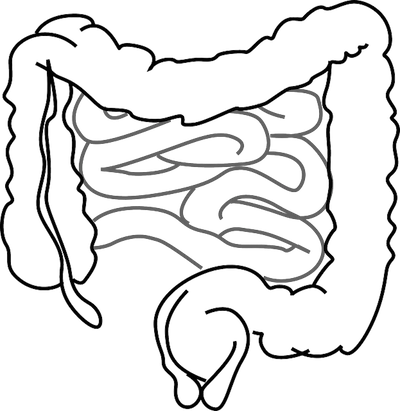
The mucus lining of our intestines are the critical interface between the microbiome and the epithelial cells that make up the intestines.  It provides many essential roles in its interaction with the microbiome, but perhaps most importantly it is a physical barrier that separates microbiome bacteria from the vasculature.  Without it, bacteria can elicit an immune response which results in inflammation that is characteristic of IBDs.  New research out of Sweden shows how mucus changes over time as the result of microbiome colonization.  The results were published in the journal Cell Host and Microbe last week.

The researchers undertook a number of experiments whereby they measured the mucus in germ free and conventionally raised mice.  In general, the conventionally raised mice had thinner, more easily shed mucus in their small intestines that allow for diffusion of nutrients.  This mucus contained antimicrobial peptides that prevented bacteria from passing across it.  The conventional mice’s large intestines’ mucus was thick and stiff and impenetrable to bacteria.  This mucus maintained most of its properties even after the conventionally raised mic were treated with antibiotics.  On the other hand, the germ free mice had thin and stiff mucus in their small intestines, as well as thick, but easily penetrable mucus in their large intestines.  This shows that the conditions under which the mucus develops is important to its eventual structure and function.  After, the scientists inoculated some of the germ free mice with the microbiome of the conventional mice and monitored the mucus over time.  It took an entire 6 weeks for the mucus to finally resemble the mucus of a conventionally raised mice.  In addition, the scientists looked that the glycans that were being formed in the mucus, and also noticed differences between the germ free and conventional mice.  As discussed on this blog previously, these glycans can be important in determining which bacteria colonize the gut.  To that end, the researchers measured the microbiome in the mice and discovered that conventional mice had a higher Firmicutes to Bacteroidetes ratio compared to germ free mice.  In addition, even after the germ free mice had been inoculated with the new bacteria, their microbiomes never truly matched the conventionally raised mice’s.

More than anything, this paper shows us the critical importance that mucus plays in microbiome health, science and research.  It demonstrates the importance of an early life microbiome to the maturation of a healthy mucus that can properly regulate the microbiome.  It also shows a possible negative consequence of antibiotics or dietary compounds can have on the mucus, and by extension the microbiome.  Finally, among many other things, it shows that microbiome research should consider the effect of the mucus on their experiments, especially ones involving germ free mice. 

Please email blog@MicrobiomeInstitute.org for any comments, news, or ideas for new blog posts.

The views expressed in the blog are solely those of the author of the blog and not necessarily the American Microbiome Institute or any of our scientists, sponsors, donors, or affiliates.

Commensal bacteria likely prevent wound healing after certain surgeries

Gastrointestinal bobbins used to connect pieces of the colon and create anastomoses

Gastrointestinal bobbins used to connect pieces of the colon and create anastomoses

Often times when people undergo colon surgeries the colon must be re-adjoined, creating what is called an anastomosis.  Around 50% of the time this anastomosis will not heal properly, and bacteria can escape from the gut into the body.  Even with improvements to surgical techniques the rates for anastomotic leak have not really improved over the years.  In fact, due to the likelihood of a patient developing this sometimes-deadly ailment, many surgeons will not even attempt to re-adjoin a colon, and instead just create a stoma (i.e. divert the colon outside the body).  It has been known that bacteria somehow cause anastomotic leaks, so intravenous antibiotics are given as standard care, but they have not been highly effective because the mechanism behind the bacterial involvement is still unknown.  However, a big step forward in elucidating the microbiome’s contribution to this disease was made by a group out of the University of Chicago.  They recently discovered that common gut bacteria, Enterococcus faecalis and Proteus mirabilis, actually hinder the bodies natural wound repair process.  They published their results in the Science Translational Medicine last week.

 First, the scientists performed a colon resection on mice and then tracked which ones developed anastomotic leaks and which ones healed normally.  Eventually they sacrificed the animals and made a number of measurements on the resectioned colons, which allowed them to compare the results between the groups.  The first thing they discovered was that enzymes responsible for degrading collagen were much higher in mice that had the leaky anastomoses. (Collagen a molecule produced by the body that is important to reattaching the sections.)  Next, they cultured organisms that were taken from the sites of the anastomoses and measured their ability to degrade collagen.  They found that two members of the communities, Enterococcus faecalis, and Proteus mirabilis, had the highest collagen degradation activity.  Interestingly, only some strains of E. faecalis had high collagen degradation activity, and it was these exact strains that were found in the leaky anastomoses, whereas the healthy anastomoses contained the other, non-collagen degrading, E. faecalis.  Moreover, when the researchers transplanted these virulent strains of E. faecalis into the guts of mice that just underwent colon sectioning these mice developed leaky anastomoses regardless of whether or not they were given antibiotics intravenously.  However, when antibiotics were applied directly to the anastomoses and E. faecalis was not able to grow, leakiness did not develop.  In a final experiment the researchers cultured bacteria from humans that had just undergone colon surgery and discovered that their anastomoses contained E. faecalis and other collagen degrading bacteria (although only one developed anostomatic leak).

 The researchers actually went a step further and pinpointed the actual genes that were associated with degrading the collagen, and I leave it to our most interested viewers to read the paper and find out more.  Thanks to research like this, and better, more targeted antibiotics, doctors are a step closer to curing leaky anastomoses.

Please email blog@MicrobiomeInstitute.org for any comments, news, or ideas for new blog posts.

The views expressed in the blog are solely those of the author of the blog and not necessarily the American Microbiome Institute or any of our scientists, sponsors, donors, or affiliates.

Understanding spatial relations of gut bacteria in ulcerative colitis patients

To sample the microbial communities in the gut, fecal samples are generally collected from an individual and DNA is sequenced to identify bacteria that are present. This is an overall effective method, however, it does not provide information of the specific spatial location of bacteria within the gut. In a study published in the journal Gut, researchers in Ireland looked to determine differences in the bacterial composition of specific regions of the large intestine between patients with ulcerative colitis and control patients.

Four volunteers undergoing routine colonoscopies were recruited to serve as the controls for this study. Five patients with ulcerative colitis (UC), who were undergoing colectomies, or surgical removal of the colon, were also involved in the study. Samples were taken at four locations in the colon in all individuals: the caecum, traverse colon, descending colon, and rectum. The four locations were sampled three times at three different levels: luminal brush, whole mucosal biopsy, and laser captured sample of mucus gel layer. A total of twelve samples were taken per individual.

After analysis of the many samples it was discovered that there was more variability between the bacterial compositions between subjects than there was within the different locations of an individual’s colon. The findings showed a difference between the luminal and mucus gel microbiota in both the controls and the ulcerative colitis subjects. Three bacterial families were common between this difference shared by controls and UC subjects, namely Bifidobacteriaceae, Peptostreptococcaceae, and Enterobacteriaceae being more abundant in UC patients.

This study has its limitations because of the small sample size, however the researchers state that the small sample allowed for extensive analysis of the individual samples. So what do the findings of this study mean for patients with ulcerative colitis? Better understanding of differences in the spatial relations of bacteria could lead to the modulation of microbial communities to help treat ulcerative colitis. 

Please email blog@MicrobiomeInstitute.org for any comments, news, or ideas for new blog posts.

The views expressed in the blog are solely those of the author of the blog and not necessarily the American Microbiome Institute or any of our scientists, sponsors, donors, or affiliates.

The fungal microbiome may be associated with colon cancer

We don’t write much about the fungal microbiome on this blog, but it may be every bit as important as the bacterial microbiome (and let’s not forget about the archaeal and eukaryotic microbiomes, and virome as well!).   Fungi are not as abundant in the microbiome as bacteria, which is probably why they are not as heavily researched, but they are known to cause diseases.  For example, vaginal yeast infections and oral thrush are caused by fungi belonging to the Candida genus.    

We recently wrote about a study that linked bacterial biofilm formation with colorectal cancer.  In this blog we mentioned that colorectal cancer is likely to have environmental causes.  Researchers from China hypothesized that fungi may be one of these risk factors, so they conducted an experiment to find out.  They recently published their results in Nature Scientific Reports.

The researchers first sampled the microbiomes of 27 patients with various stages of colorectal tumors, in addition to other, healthy areas of those patients’ guts adjacent to the tumors.  They then sequenced the genomes of the samples to determine which fungi existed, and where.  They discovered that fungal diversity was lower on tumors compared to other areas of the colon.  In addition, two known pathogenic fungi, Candida and Phoma existed in higher levels on tumors compared to the adjacent areas.  Finally, they found distinct differences between individuals with advanced and non-advanced tumors.  Those with advanced tumors had a higher abundance of two other known pathogenic fungi, Fusarium, which has been associated with intestinal disease in the past, and Trichoderma, which has been associated with infections of various organs. 

This study did not involve any healthy patient controls, and its sample size was somewhat limited.  Still, the results are intriguing because gut fungi that are known to cause inflammation elsewhere in the body are being found at the site of tumors.  Even if these fungi are not causing the tumors, they could at least be potentially used as a diagnostic or biomarker for tumors.  While we know that some fungi can be dangerous, we note that even specific genera are not always pathogenic, and sometimes they can exist normally in a host and only turn pathogenic at a later time.  Like other aspects of the microbiome, the story is complicated, but we would be willing to bet there is at least one beneficial fungus among us.

Please email blog@MicrobiomeInstitute.org for any comments, news, or ideas for new blog posts.

The views expressed in the blog are solely those of the author of the blog and not necessarily the American Microbiome Institute or any of our scientists, sponsors, donors, or affiliates.

The effects of bowel cleansing for colonoscopies on our microbiome

Colonoscopies and colonic surgeries are known to require bowel cleansing prior to the procedure.  To accomplish this, what are thought to be generally safe and effective products are used in order to cleanse the bowel. One commonly used cleanser is polyethylene glycol (PEG) supplemented with electrolytes. While the efficiency of this product has been tested, limited data has been collected on its effects on the microbiome of the colon.

A new study published on January 6th in the journal Gut tested the effects of bowel cleansing on the bacteria that colonize an individual’s gastrointestinal tract. 23 human subjects were put into two study groups, one group consuming the PEG electrolyte solution in a single dose of 2 L and the other group consuming the solution in two doses of 1 L.

They found that bowel cleansing had little long-term effect on an individual’s colonic microbiota though a substantial change was initially seen after consumption. In subjects who consumed the PEG electrolyte solution in two 1 L doses, the microbiota recovered to original composition after two weeks. In subjects who consumed the solution in a single 2 L dose, however, it took up to one month after treatment for the colonic microbiome composition to recover.

Due to these findings, the authors suggest that separating the dosage into two 1 L doses may benefit the microbiome of the patient and may be preferred in clinical practice. There are several new microbiome studies such as this one that could impact medical practice and it will be exciting to see how (and if) the outcomes outlined in these papers are adopted by clinicians into practice.  

Please email blog@MicrobiomeInstitute.org for any comments, news, or ideas for new blog posts.

The views expressed in the blog are solely those of the author of the blog and not necessarily the American Microbiome Institute or any of our scientists, sponsors, donors, or affiliates.

Microbiome based therapy prevents weight gain in obese individuals

New microbiome based drug may cause people to eat less!

New microbiome based drug may cause people to eat less!

Many people have heard that eating fiber is good for your health and helps to prevent weight gain.  One of the reasons for this, as we have blogged about before, is thought to be related to the short chained fatty acids (SCFAs) that are produced by the microbiome from fiber.  In mouse models, mice that receive a fecal transplant containing a microbiome with a high capacity for generating SCFAs show reduced weight gain.  The SCFAs appear to induce the production of certain hormones associated with appetite control.  The problem with human interventions of fiber-based diets is that an unpalatable amount of fiber is normally required, and very little of it is eventually converted into SCFAs.  Scientists from England recently tried to tackle this problem by introducing one type of SCFA, propionate, into the colons of obese humans to investigate its effects on weight gain.  The results of their study were published in the journal Gut.

The researchers chemically modified propionate so that after it was eaten it would only be released in the colon.  They then performed a double blind trial with 60 obese participants that took either the propionate or a placebo every day for 24 weeks.  The scientists discovered that, as hypothesized, ingesting the propionate increased the production of the appetite control hormones in the colon.  In addition, people who took the propionate tended to have a suppressed appetite and ate less overall food than their placebo counterparts.  Overall, the propionate prevented weight gain in the individuals who ingested it compared to the placebo group.

Recent advances in microbiome research have shown hormones produced by the gut are critical to managing hunger and food intake, and that research has allowed these scientists to create a new drug that stimulates the microbiome into producing those hormones.  In addition, the scientists show a new method of drug delivery to the colon which may have applications for other therapies.  At the AMI, we hope that their research continues to be as fruitful as it is promising.

Please email blog@MicrobiomeInstitute.org for any comments, news, or ideas for new blog posts.

The views expressed in the blog are solely those of the author of the blog and not necessarily the American Microbiome Institute or any of our scientists, sponsors, donors, or affiliates.