plos one

The oral microbiome of children, and its relation to dental caries

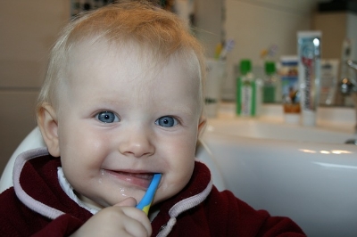
The oral microbiome is a popular area of exploration because bacteria are a prominent part of dental health, and because it is one of the most heavily colonized and easily accessible niches in the body. Many studies have been discussed on this blog concerning adult oral microbiomes, and its relations to bodily issues such as cystic fibrosis and periodontitis. It is also very useful to investigate children and the ways that their bacterial communities first inhabit and develop. A study done in Sweden at the Umeå University, and published by Plos One, takes a look at the maturation of the oral microbiome from infants at 3 months old to children at 3 years old.

The Swedish researchers performed a longitudinal study that followed children from 3 months to 3 years of age, looking for microbial characteristics of children with dental caries (i.e. cavities) compared to those without. There were 207 original participating 3 month olds that were consented by their parents to be in the study. The parents provided information on mode of feeding, mode of delivery, use of antibiotics or probiotics, health issues like allergies, and presence of teeth. At 3 months and later at 3 years samples were taken from the buccal mucosa, tongue, and alveolar ridges. Teeth were also scraped for plaque and saliva was collected. Of the original 207 participants, 155 returned for sampling at 3 years of age, and 13 of those children had dental caries.

After sequencing the bacterial DNA samples, it was found that Escherichia coli, Staphylococcus epidermidis, and various Pseudomonas species were significantly more prevalent in 3 month olds. However, there were 23 genera that were more significantly prevalent at 3 years of age than at 3 months.

By comparing the children with and without caries, the scientists were able to make several conclusions.  The researchers identified seven taxa that appear to be associated with healthy teeth.  On the other hand, Streptococcus mutans seemed to be more prevalent in the children with caries, than in those without caries. Additionally, the colonization of this species was most prevalent in girls. This is possibly because girls develop faster, so earlier tooth eruption allows for a longer time for the colonization of these bacteria.

The results of this study show us that during the first three years of life, species richness and diversity seems to increase significantly in the mouth. While there is an increase in the type of the bacteria, there are also some taxa that are lost with age. The researchers also concluded that the oral microbial composition of the mouth at 3 months does not appear related to the development of dental caries. With this information, it might be smart to perform a related study that collects oral microbiome samples in children within the time frame of 3 months to 3 years, because it could show a clearer picture of the changes that take place in bacterial composition.

Please email blog@MicrobiomeInstitute.org for any comments, news, or ideas for new blog posts.

The views expressed in the blog are solely those of the author of the blog and not necessarily the American Microbiome Institute or any of our scientists, sponsors, donors, or affiliates.

Common yeast may trigger celiac disease onset

Candida albicans growing in petri dish

Candida albicans growing in petri dish

Celiac disease is a serious autoimmune disorder in which gluten, found in wheat, rye, and barley, triggers an immune response that damages the absorption capabilities of the intestines. The AMI has covered this topic in a few previous blogs (click celiac disease tag below) in relation to autoimmune disorders and possible bacterial triggers. One contributing factor to the celiac disease response is due to the protein gliadin, which is found in gluten. Gliadin, along with transglutaminase (which is a human protein that binds to, and deaminates gliadin), trigger a T Cell response that leads to the inflammation and tissue damage.

The yeast Candida albicans is a common gut commensal that is linked to inflammatory bowel diseases and vaginal infections.  This yeast also binds with transglutaminase, using a protein called Hwp1, in an identical fashion as gliadin.  This results in the bacteria’s strong binding to the intestinal wall, where it triggers an autoimmune response to destroy the yeast.  

Researchers in France hypothesized that the similarity between gliadin's and C. albicans' binding to transglutaminase may result in a similarity in the body's response to these two things.  In essence, they suggested that gluten ‘tricks’ the body into an immune response because it 'looks' similar to C. albicans.  

In the study, recently published by Plos One, blood cultures from 87 adult patients with celiac disease and 41 patients with C. albicans infection were collected.  The scientists then isolated the body's natural antibody for Hwp1 and measured its response to both gliadin and Hwp1.  They discovered that gliadin also binds to Hwp1's antibody, meaning that it should elicit the same immune response as Hwp1.  Therefore, the body should mount an immune response for gluten that is characteristic of C. albicans infection, and this response could manifest itself as celiac disease.

The significance of this study is that it comes closer to finding a cause and prevention of celiac disease. The T cell immune response that results from transglutaminase binding to gliadin could initially be triggered by a C. albicans yeast infection. This may explain why some people only become gluten sensitive later in life - perhaps it only occurs after they have a C. albicans infection and the body builds up antibodies for this yeast. This is another example of how microbes found in healthy individuals can be harmful when homeostasis is not controlled. 

Please email blog@MicrobiomeInstitute.org for any comments, news, or ideas for new blog posts.

The views expressed in the blog are solely those of the author of the blog and not necessarily the American Microbiome Institute or any of our scientists, sponsors, donors, or affiliates.

The blood microbiome and infection after transfusion

Red blood cells in plasma

Red blood cells in plasma

In general, a significant amount of attention is placed on microbiome communities in our gut and skin, and their respective relationships with other tissues in our bodies (the brain, for example).  However, the cells and fluids that are essential to our survival, such as blood, may also play an important interactive role with our microbiome.  This relationship is demonstrated in a recent study aimed at identifying bacteria found in standard blood-packs used for blood transfusions, and examining bacterial distribution and distinct strains present in blood-plasma and red blood cells. 

Transfusion-transmitted infection remains the leading cause of post-blood transfusion mortality and morbidity even though these risks have declined significantly in recent years.  As the name suggests, transfusion-transmitted infections result from the introduction of foreign pathogens to a patient’s blood stream via a blood transfusion.  However, previous research has identified a significant discrepancy between post-transfusion infection rate and bacterial growth observed in the blood pack from which the transfusion was received.  Specifically, a 16.9% rate of post-transfusion infection (11.8% under more reserved transfusion methods) is observed.  However, data from standardized bacterial screening protocols indicate that less than 0.1% of blood packs actually contain bacterial growth. 

Previous literature examining bacteria translocation into red blood cells concomitant to epidemiology data related to gum disease suggest that the oral cavity, or mouth, can serve as a viable access point for bacteria to enter into the blood stream.  Furthermore, conventional methods used to screen for bacteria presence in blood packs does not account for bacterial adherence to red blood cells.  To address this discrepant data and literature-supported suppositions, a twofold approach was taken.  Researchers sought to determine if known oral cavity bacteria strains are found in donor blood packs and whether or not these bacteria adhere to red blood cells.

Blood was drawn from 60 healthy study participants and subjected to specific fractionation procedures to separate red blood cells from plasma.  Red blood cell and blood plasma suspensions were subsequently plated on cell culture dishes and incubated for 7 days under specific conditions to allow researchers to isolate bacteria from red blood cells and identify the strains.  General bacterial growth was evident in both red blood cell and blood-plasma dishes.  Of the 60 plates corresponding to 60 patients, marked growth was observed in 35% of the red blood cell cohort and 53% of the blood-plasma cohort.  DNA amplification of known bacteria found in the oral cavity was then used to determine the specific bacterial strains that were incubated on these plates.  Various aerobic and anaerobic strains were identified and it was interestingly noted that these bacteria are undetectable using standardized bacterial screening techniques.

These findings certainly have major implications for clinical diagnosis of bacterial contaminants found in blood packs.  In particular, detection capabilities of diagnostics must be improved, as it turns out that there may be significant amounts of bacteria in blood packs than previously realized.  This study also illustrates that bacterial communities are mobile and are not limited to gut, skin, mouth, vagina, and other tissues we normally associate with the microbiome.  The ability to adhere to red blood cells certainly gives microbiota populations a much more dynamic range of influence in our body’s respiratory, immunologic, and overall regulatory processes.  

Please email blog@MicrobiomeInstitute.org for any comments, news, or ideas for new blog posts.

The views expressed in the blog are solely those of the author of the blog and not necessarily the American Microbiome Institute or any of our scientists, sponsors, donors, or affiliates.

The microbiome of alcoholics may contribute to pathologies

We have written before about the microbiome’s association with alcoholism, and how it has been implicated in many of the maladies connected with the disease.  Recently, research out of George Mason University, published in PLoS ONE, explored the molecular mechanisms behind this relationship.   The scientists measured the metabolites that were formed by the microbiome of alcoholics and compared it to healthy controls.  They discovered that the metabolites that differed between the two groups have important implications on gut health.

The scientists measured the volatile molecules that were being effused from the feces of 18 healthy controls and 16 alcoholics.  The alcoholics’ feces contained high levels of an organic compound called tetradecane, which is known to cause oxidative stresses.  Increased oxidative stress in the gut, especially in alcoholics, is associated with increased gut permeability (i.e. leaky gut), and alcoholic steatohepatitis (i.e. a type of liver disease).  Moreover, specific fatty acids, which are known to reduce oxidative stress (antioxidants), were more depleted in alcoholics when compared with healthy controls.  In addition, the alcoholic feces consisted of lower abundances of short chained fatty acids (SCFAs), which are nearly always associated with intestinal health (click the SCFA tag below to learn more).  Finally, other molecules which are associated with health, like caryophyllene and camphene, were decreased in the guts of alcoholics.

Overall these results show the possible mechanisms by which the microbiome contributes to alcoholism.  Specifically, it appears that the alcoholic microbiome may create oxidative stress molecules, which contribute to gut toxicity.  In addition, the scientists suggest this work could be used as an alcoholism diagnostic, as the characteristic metabolites between the groups were statistically significant.


Please email blog@MicrobiomeInstitute.org for any comments, news, or ideas for new blog posts.

The views expressed in the blog are solely those of the author of the blog and not necessarily the American Microbiome Institute or any of our scientists, sponsors, donors, or affiliates.

Walk, chew gum, and fight cavities all at the same time

Chewing gum has been suggested to have many cognitive benefits such as increasing focus and alertness, improving memory, and controlling, besides its obvious benefit of making breath smell minty fresh.  What about gum's effect on our microbiome though?  Could gum help prevent cavities in the same way as tooth brushing or flossing?  In order to find out, researchers from the Netherlands and Wrigley, the gum company, recently published the results of a study in PLoS One that set out to answer the question: how much oral bacteria is trapped and removed by chewing gum?

In order to test the hypothesis, volunteers chewed gum for various times for up to 10 minutes. The researchers then used different quantitative and qualitative analyses, such as culturing and genomic analysis, to measure the amounts of bacteria collected in the gum.  The researchers found that the chewing gum does indeed trap around 100 million bacteria, which is about the same as brushing your teeth with a new, clean toothbrush without using toothpaste. They also state that chewing gum could prevent biofilm formation, much like tooth brushing.  Finally, they concluded that the longer gum is chewed, the fewer bacteria it removes from the mouth.

This study in quantifying bacterial removal by gum was preparing the researchers for their next project, which is to intelligently design gum to prevent cavities.  As we know there are healthy and harmful bacteria in the oral cavity, but the study did not investigate which types of bacteria were removed.  If gum could be designed that preferentially adsorbs and removes acid-forming bacteria like Streptococcus mutans then it could be highly effective in eliminating cavities.  We look forward to reading more about this project, and in the meantime, if you’re going to chew gum, try and make sure it’s sugar free.

Please email blog@MicrobiomeInstitute.org for any comments, news, or ideas for new blog posts.

The views expressed in the blog are solely those of the author of the blog and not necessarily the American Microbiome Institute or any of our scientists, sponsors, donors, or affiliates.

Asthma, COPD, and the Microbiome

Asthma and chronic obstructive pulmonary disease (COPD) are both illnesses that are caused by chronic inflammation of the respiratory tract, and recent research suggests that the microbiota of the lower respiratory tract may influence the development of these two diseases.  The upper respiratory tract, though, remained unstudied, until a new article was recently published in PLoS ONE.  This article characterized the microbiome of the oropharynx (in the upper respiratory tract) to discover the association between these problems and the microbiome.

Samples were swabbed from the oropharynx of patients who were recently diagnosed with asthma and COPD, as well as from a healthy control group.  Researchers performed 16S rRNA gene sequencing of the bacteria collected from the patients, in order to determine which bacteria were present. They found that there are few differences in microbiome diversity between asthma and COPD patients, however there was a prevalent presence of the bacteria Lactobacillus (phylum Firmicutes) and Pseudomonas (phylum Proteobacteria) in both, which were identified in only very small amounts in healthy patients. On the contrary, the upper respiratory tract of healthy individuals was found to be dominated by Streptococcus, Veillonella, Prevotella, and Neisseria, from the phylum Bacteroidetes, compared to individuals with asthma and COPD.

This study showed distinct differences in the microbiomes of diseased and healthy individuals.  The researchers also note that the low abundance of Neisseria they observed in this study has also been seen in studies of smokers, meaning that this bacteria may be important to respiratory health.  Further work is still needed, though, to determine if the bacteria identified in this study are contributing to the diseased individuals.  Even if they are not, they could still potentially be used in diagnosis. 

Please email blog@MicrobiomeInstitute.org for any comments, news, or ideas for new blog posts.

The views expressed in the blog are solely those of the author of the blog and not necessarily the American Microbiome Institute or any of our scientists, sponsors, donors, or affiliates.