Gut bacteria may prevent kidney injury

Scientists have found that short chain fatty acids (SCFAs), a product of gut bacteria, may protect the kidneys from acute kidney injury (AKI), a condition with high mortality rates that can also lead to other very serious kidney diseases. AKI is often caused by something called ischemia reperfusion injury, an injury resulting from a loss of oxygen to the tissue (ischemia) and a rush of blood back to the site (reperfusion). This instigates a cascade of events resulting in several immune cell populations accumulating at the site of the injury, causing inflammation and kidney damage.

Because AKI is a result of inflammation and because SCFAs are known to have anti-inflammatory effects, scientists in Brazil hypothesized that treatment with SCFAs could ameliorate kidney function. The results published in the Journal of the American Society of Nephrology were the first to show the protective role of SCFAs in kidney ischemia reperfusion injury (IRI). They found that when the three main SCFAs (acetate, propionate, and butyrate) were administered to mice undergoing this IRI injury, they protected the kidney from undergoing AKI.  As suspected, the SCFAs prevented an autoimmune response which resulted in less inflammation and apoptosis (cell death).

Acetate was the SCFA that was most protective to the kidney, so in another experiment the scientists administered acetate-producing bacteria to the mice.  Bifidobacterium adolescentis and Bifidobacterium longum were administered separately and each did produce acetate, as evidenced by increased acetate levels in the mice's feces. They found that these mice were protected from kidney IRI and therefore the bacteria were effective. They did note, though,  that it is unlikely the bacteria colonized the gut, so further investigation is needed.  

This study provides another example of probiotics preventing conditions that may have resulted in serious injury and even death. The bacteria in this study are already used in probiotics to treat other diseases, and so repurposing them for kidney disease should be possible.  The study also describes the anti-inflammatory effects of SCFAs, which we have written extensively about in this blog

Please email blog@MicrobiomeInstitute.org for any comments, news, or ideas for new blog posts.

The views expressed in the blog are solely those of the author of the blog and not necessarily the American Microbiome Institute or any of our scientists, sponsors, donors, or affiliates.

Gut microbiota reveal better ways of detecting (and preventing) colorectal cancer

Editors note:  The AMI is thrilled to have a guest blog post today from Kristina Campbell, a very talented science writer who specializes in the microbiome.  She writes for the Gut Microbiota for Health Experts Exchange, and blogs as The Intestinal Gardener.  Our readers can also keep up with her on Twitter,  @bykriscampbell.  


By Kristina Campbell

Scientists are starting to develop an idea of how colorectal carcinoma (CRC) arises. It all starts with bacteria in the digestive tract: possibly a strain of Bacteroides fragilis or the infamous Escherichia coli. Whether the bacteria are new to the body, or resided there all along, doesn't matter. The bacteria somehow get a green light to start producing chemical agents that damage the genetic information in the body's cells. The damaged cells rapidly divide. Soon enough, polyps (also called adenomas) appear in the colon. These polyps can become cancerous.

Currently, there's a gap in CRC detection methods. This is a problem that's directly connected with patient mortality: if CRC is detected at an early stage, survival rate is more than 80%. But if it's left until a late stage, it's less than 10%.

The two standard ways to detect this cancer are a fecal occult blood test (FOBT) and a colonoscopy. FOBT – which tests for traces of blood in the stool – has limited sensitivity for CRC. It's only a rough guide, since it misses many cases. Colonoscopy is the most effective method of diagnosis, but it's far from perfect because it's invasive and costly.

New research shows that the microbiota might lead to better CRC detection. Iradj Sobhani and colleagues recently published an intriguing paper in Molecular Systems Biology called ‘Potential of fecal microbiota for early-stage detection of colorectal carcinoma'. They took fecal samples from healthy people and those with confirmed CRC, and used metagenomic sequencing to find out how they differed.

They found that the fecal samples held clues that were missing from FOBT. Using both methods together, they increased the sensitivity of colon cancer detection 45% (as compared with FOBT alone). Used effectively in the clinic, this could save thousands of lives each year.

Sobhani said he and his colleagues are working on a clinical tool to help patients make use of this information. In a recent interview, he said, "Now we know a panel of some 18-20 [relevant] bacteria and we are trying to make an easy and simple tool to identify these bacteria. We can, I hope, in a very short future time, make low-cost tools to identify the bacterial phenotype usually found in patients with colon cancer."

A smaller study from the Schloss lab found a similar result: enhanced CRC detection using information from FOBT and a fecal sample, as well as body mass index, age, and race (which are known risk factors for colon cancer).

Schloss said that one kind of bacteria in particular piqued his interest. "We’re trying to better understand [why] Fusobacterium seems to be popping up in a lot of these stories. How does Fusobacterium get from the mouth to the gut? Everybody has it in their mouth. But not everybody has it in the gut. So what’s breaking down there? Does it have a role in disease?"

The Sobhani study went beyond CRC detection to factors that might be involved in prevention. The researchers looked at the bacterial genes in the guts of those with CRC and asked, "What can these bacteria do well?" In other words, they looked at the bacterial functions as indicated by their genes.

This analysis showed some interesting links to diet. Sobhani explained, "Those with colon cancer had largely more meat-metabolizing bacteria] compared to those who have no colon cancer, who have bacteria that show more functions to metabolize vegetables." He added, "Then there are functions involved in the transfer and capture of… minerals."

Whatever made the meat metabolizers more abundant in the colon could turn out to be what caused the cancer in the first place. But it's not clear whether red meat consumption itself accounts for the disease-associated condition of the microbiota, or whether other components of the diet play a role. (Fiber is a prime preventative candidate under investigation.)

A whopping 95% of CRC could be attributable to environmental factors. More research related to the gut microbiota and CRC might one day reveal exactly what those environmental factors are, so we can kick colon cancer to the curb.

References:

Zackular J, Rogers M, Ruffin M and Schloss P. (2014) The Human Gut Microbiome as a Screening Tool for Colorectal Cancer. Cancer Prevention Research doi: 10.1158/1940-6207.CAPR-14-0129

Zeller G, Tap J, Voigt AY, et al. (2014) Potential of fecal microbiota for early-stage detection of colorectal cancer. Molecular Systems Biology doi: 10.15252/msb.20145645

Please email blog@MicrobiomeInstitute.org for any comments, news, or ideas for new blog posts.

The views expressed in the blog are solely those of the author of the blog and not necessarily the American Microbiome Institute or any of our scientists, sponsors, donors, or affiliates.

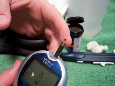
The infant microbiome changes before the onset of type 1 diabetes

file0001403630450.jpg

Type 1 diabetes (T1D) is a disease in which your immune system attacks and destroys your insulin-producing cells.  There is a known genetic risk factor in developing T1D, but there are also significant non-genetic components to getting the disease.  Previous research in mice has established the microbiome's connection with the development of diabetes, but the link in humans has not been studied as closely.  Researchers from various institutions in the U.S. and Finland recently assembled a cohort of infants genetically at-risk for diabetes, and tracked the changes in their microbiomes.  They discovered that the microbiomes of those individuals that were eventually diagnosed with diabetes underwent characteristic shifts leading up to diagnosis, and that these changes were not observed in healthy infants.  They published the results of their study in Cell Host and Microbe.  

The researchers sampled the stools of 33 infants in Finland and Estonia that were genetically at-risk for diabetes.  Their first major discovery was that even though the bacterial composition of the microbiome grew, changed, and became more diverse with age, the types and number of genetic pathways that were expressed by the microbiome, as well as the metabolites produced by the microbiome remained stable.  They also found many similar bacterial species between infants, however these infants usually had different strains of said bacterial species.  In most of these cases, once a particular strain established itself in the gut it remained stable and would not be displaced.

The scientists tracked the microbiome changes that occurred with diet as well.  During breast feeding Bifidobacterium and lactobacillus predominated, and Lachnospiraceae decreased.  After cessation of breast feeding the addition of eggs barley and soy seemed to have a direct influence on the microbiome.  One of the biggest factors in the developing microbiome was actually geography, as the Estonian infants had significantly higher levels of Bacteroides and Streptococcus species.

The researchers then compared the microbiome samples between those infants that were eventually diagnosed with diabetes and those that were not.  They discovered that a few bacterial species were much more abundant in those infants that got diabetes: Blautia, the Rikenellaceae, and the Ruminococcus and Streptococcus genera, including Ruminococcus gnavus and Streptococcus infantarius.  Interestingly, each of these bacteria are ‘pathobionts’, or bacteria which exist in many healthy peoples’ microbiomes but have the potential to become pathogenic.  Also, certain bacteria such as Coprococcus eutactus and Dialister invisus were non-existent in the diabetics' guts.  In addition, the researchers discovered that the expression of specific genes, like those associated with sugar transport and the biosynthesis of amino acids, underwent shifts prior to the onset of diabetes.  Finally, many of these bacteria that were associated with T1D appeared right before the onset of the disease, and these bacteria were linked to the presence and absence of certain metabolites in the stool.

These results provide exhaustive evidence for an association between the microbiome and diabetes.  It links specific bacteria in the microbiome and the expression of certain genes by the microbiome to the disease.  The next step is to study the mechanisms by which the microbiome induces diabetes, and then therapeutics can be developed.


Please email blog@MicrobiomeInstitute.org for any comments, news, or ideas for new blog posts.

The views expressed in the blog are solely those of the author of the blog and not necessarily the American Microbiome Institute or any of our scientists, sponsors, donors, or affiliates.

What does NYC's microbiome look like?

Grand Central Station

Grand Central Station

Every week we talk about the human microbiome that makes up each and every one of us, but what about the environmental microbiome, particularly in urban areas?  Cities are just like people (kind of), and they have a microbiome too! A very cool study published yesterday in Cell Systems describes New York City's microbiome.  A large team of scientists, led by a group at Weill Cornell Medical College investigated the DNA profile of the city’s public transit system, several public spaces, and the Gowanus Canal.

They collected samples from every open NYC subway station (466 of them), the Staten Island Railway, 12 locations along the Gowanus Canal in Brooklyn, four public parks, as well as the South Ferry Station that was closed at the time of sampling due to flooding from Hurricane Sandy. At the subway stations, they sampled inside the trains, kiosks, benches, turnstiles, garbage cans, and railings. The results were fascinating. 

Almost half of all the reads could not be matched to any known organism.  They found that South Ferry Station, the only station in NYC that was completely submerged in Hurricane Sandy, represented a marine environment.  They discovered that some known pathogenic bacteria, such as Yersinia pestis, the cause of the bubonic plague, and Bacillus anthracis, anthrax, were present in a few subway stations.

Not surprisingly, the scientists found that the most common bacteria in these locations were those that are abundant on humans' skin.  This makes sense as thousands of people pass through each one of the subway stations every day.  In addition, stations with the greatest levels of traffic on a daily basis, like Grand Central and Times Square, had the greatest diversity of bacteria.

What about all these unknown organisms that couldn’t be identified? These exemplify the huge amount of life right in front of us that has yet to be characterized.  What about all the pathogenic bacteria and antibiotic resistant bacteria like MRSA that were identified? Should we be worried that these? The authors state that the subways generally contain safe surfaces for the following reasons. First, they note that it is impossible to tell if the sequenced organisms are dead or alive. Second, even more importantly, these bacteria help make up a dynamic and shared urban infrastructure, and they may even help maintain its equilibrium. Finally, the bacteria that were connected to the plague, anthrax, and other diseases were found in very small traces, not enough to colonize our bodies. (Did you know that people in the United States, almost exclusively on the West Coast, are still getting the bubonic plague today?)

It will be very interesting to see what comes from this study. I, for one, would like to know what the South Ferry Station microbiome looks like now compared to when the scientists initially sampled, because the sampling was performed when it was still closed due to Hurricane Sandy.  Now that it is back up and running with thousands of people passing through it every day, how has the ecosystem of the station changed?. 

The authors suggest that a study like this could help facilitate forensic investigations. For example, the bottom of one’s shoe could represent where that person traveled over the past day or week. They also suggest that faster, or maybe even real-time analysis, of the microbiome of the city’s transit system could be used to protect the city from bioterrorism and other public health threats like our recent scare with Ebola. 

If you want to see what bacteria were found at your favorite station, The Wall Street Journal put together a very easy-to-use interactive map. Check it out here.

Please email blog@MicrobiomeInstitute.org for any comments, news, or ideas for new blog posts.

The views expressed in the blog are solely those of the author of the blog and not necessarily the American Microbiome Institute or any of our scientists, sponsors, donors, or affiliates.

Research is needed on the role of the microbiome and anorexia nervosa

Sketches of Miss A before (top) and after (bottom) treatment for anorexia nervosa by William Withey Gull.  Dr. Gull was among the first people to be clinically establish and treat anorexia, and was responsible for naming the disease.

Sketches of Miss A before (top) and after (bottom) treatment for anorexia nervosa by William Withey Gull.  Dr. Gull was among the first people to be clinically establish and treat anorexia, and was responsible for naming the disease.

Yesterday we discussed the latest research on the microbiome and obesity.  Today we wanted to share some thoughts on the other end of the spectrum: the microbiome and anorexia nervosa.  This week in the International Journal for Eating Disorders there was a call for research by doctors from the University of North Carolina to investigate the microbiome’s role in this disease.

Anorexia nervosa is a neurological disease characterized by self-starvation.  It often occurs with depression and other affective disorders.  It has severe side effects such as heart disease and seizures, which makes it the deadliest neurological disease, with around 5% of cases becoming fatal.  The causes of anorexia are complex, but genetic, social, and environmental risk factors all exist. 

Anorexia nervosa is a disease that is very likely implicated with the microbiome, perhaps profoundly.  It coincides with our understanding of the so-called gut-brain axis, which has already been linked to depression, anxiety, and appetite, which are distinctly awry in anorexia sufferers.  The relationship between anorexia and the microbiome is largely unknown.  Studies have indicated those suffering from anorexia have very unique microbiomes with similar bacterial that are not commonly found in the gut, but these studies are hardly convincing and not robust. 

Treatment for anorexia always includes dietary interventions, but these are not always effective in reestablishing normal weight, and relapse occurs in around half of all patients.  Recovery and therapeutics should also consider the microbiome.  Research on malnourishment in Africa has produced a wealth of literature on the importance of a balanced, robust, and healthy microbiome to nutrition and weight.  Simply adding calories to a diet is often not enough to improve health in the malnourished because their dysbioses, and the same phenomenon may also occur in patients suffering from anorexia nervosa.

Because the microbiome may be fundamental to both the neurological and dietary aspects of the disease, the authors of the paper encourage further research.  They suggest that microbiome transplants may be important to both physical and mental healing.  Anorexia nervosa is a complex disease, but research on the microbiome is crucial to fully understanding it.

Please email blog@MicrobiomeInstitute.org for any comments, news, or ideas for new blog posts.

The views expressed in the blog are solely those of the author of the blog and not necessarily the American Microbiome Institute or any of our scientists, sponsors, donors, or affiliates.

New evidence for the relationship between obesity and the microbiome

Obesity is rapidly becoming a big problem in the United States (no pun intended), and up to one-third of adults are obese, according to the Centers for Disease Control and Prevention. As a disease, obesity is not well understood because there are so many factors that are involved in weight control, including caloric intake, genetic predisposition, and psychological factors among many others. A study done by researchers at the Institute of Nutritional Sciences, of the University of Vienna, and recently published by Beneficial Microbes, considers another factor involved in obesity: the microbiome.  Previous research on mice has shown that the microbiome is associated with obesity and leanness, and that diet can be an important tool to modulate it.  These researchers extended some of these ideas to humans, and studied the microbiomes of obese people as they lost weight through diet and exercise.

The researchers studied 33 obese participants during a four month period of strict diet and exercise regime, which resulted in weight loss. Fecal samples were collected once before, during, and after the study and the bacterial content was tested.

A decisive difference in the bacterial content of the gut was found in individuals before the study and after weight loss. Significant findings of this study include the increased presence of Lactobacilli after weight loss, a genera that has been found to have probiotic, anti-inflammatory characteristics.  The study also found that weight loss was associated with a decrease in the ratio of Firmicutes to Bacteroidetes, with the abundance of Firmicutes increasing in presence and the abundance of Bacteroidetes remaining the same.  Interestingly, the amount of weight lost was related to the amount decrease in this ratio.  Finally, the scientists found that Archaea, which are a distinct phylogenetic domain from Bacteria and Eukaryotes, increased in abundance after weight loss.

As we know, the microbiome holds a lot of promise in the fight against obesity, and this study, as well as those that are sure to follow, could impact obesity treatments in the future.  We need to remember, though, that far more rigorous studies are needed before any actionable strategies to modulate the microbiome to lose weight are established.  Perhaps though, the key to achieving a healthy weight is to maintain a certain level of bacterial diversity in the gut.

Please email blog@MicrobiomeInstitute.org for any comments, news, or ideas for new blog posts.

The views expressed in the blog are solely those of the author of the blog and not necessarily the American Microbiome Institute or any of our scientists, sponsors, donors, or affiliates.