amino acids

Maternal stress can alter the gut microbiome of progeny, possibly affecting brain development

The composition of the vaginal microbiome has been shown to have major health implications for a female’s health as well as the health of a newborn infant.  During birth, microbiota transfer from the mother to the neonate, which eventually go on to colonize the gut of the child.  It has already been shown that disruptions to the vaginal microbiome can impact microbiota colonization in the gut of a neonate, but downstream implications of this have not been thoroughly explored. 

Researcher’s from University of Pennsylvania set out to examine whether maternal stress in mice, and subsequent changes to the vaginal microbiome, could lead to disruptions in the gut microbiome of their progeny.  Expanding upon this, the researchers further investigated whether these disturbances to the gut impaired metabolism.  This transfer of microbiota occurs during a critical time in brain development, which requires a lot of energy and therefore effective metabolism to fuel this process.  The researchers wanted to identify whether or not maternal stress could disrupt the brain development process by way of alterations to microbiome transfer from the mother to its progeny and a subsequent disrupted metabolic process. 

Male C57 mice and female 129S1 mice were used in this study and were bred to form a hybrid F1 generation.  Stress was administered to the female mice using a well-established behavioral paradigm known as the early prenatal stress model.  Pregnant mice assigned to the EPS-stress group were exposed to a series of stressors (8 in total), but pain was not induce nor did these tests directly influence feeding schedule, weight gain, and litter size. 

Animals were then sacrificed and vaginal lavages were collected to examine bacterial composition between stressed (EPS) and non-stressed groups.  Quantitative PCR was used to characterize the microbiomes of the female mice and their offspring.  Lactobacillus, the predominant bacteria populations in the vagina, was significantly disrupted in the EPS group.  There was a reduction in Lactobacillus in the guts of F1 progeny as well.

Colon and plasma metabolic samples were examined in the F1 hybrid generation by extracting fatty acid metabolites using centrifugation.  Analysis showed that metabolic profiles were significantly different between groups.  Namely, of 29 signature metabolites assessed, 6 were increased and 23 were decreased in EPS progeny as compared to the control groups. 

Brain samples of the F1 hybrid generation were collected and amino acid concentrations were analyzed to assess substrate availability in the developing brain.  The F1 offspring from the EPS group displayed significantly less amino acids.  Interestingly, amino acids in a hypothalamic region of the brain were shown to be deregulated, and these concentrations were much lower in males as compared to females. 

It was interesting to see differences in amino acid availability in the hypothalamus between males and females in light of the fact that there are gender biases in neurodevelopmental disorders such as autism spectrum disorder.  Hopefully future studies can elucidate more on the microbiome to see how it relates to human behavior and brain disease.  

Please email blog@MicrobiomeInstitute.org for any comments, news, or ideas for new blog posts.

The views expressed in the blog are solely those of the author of the blog and not necessarily the American Microbiome Institute or any of our scientists, sponsors, donors, or affiliates.

The infant microbiome changes before the onset of type 1 diabetes

file0001403630450.jpg

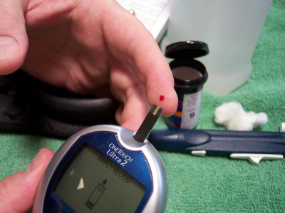
Type 1 diabetes (T1D) is a disease in which your immune system attacks and destroys your insulin-producing cells.  There is a known genetic risk factor in developing T1D, but there are also significant non-genetic components to getting the disease.  Previous research in mice has established the microbiome's connection with the development of diabetes, but the link in humans has not been studied as closely.  Researchers from various institutions in the U.S. and Finland recently assembled a cohort of infants genetically at-risk for diabetes, and tracked the changes in their microbiomes.  They discovered that the microbiomes of those individuals that were eventually diagnosed with diabetes underwent characteristic shifts leading up to diagnosis, and that these changes were not observed in healthy infants.  They published the results of their study in Cell Host and Microbe.  

The researchers sampled the stools of 33 infants in Finland and Estonia that were genetically at-risk for diabetes.  Their first major discovery was that even though the bacterial composition of the microbiome grew, changed, and became more diverse with age, the types and number of genetic pathways that were expressed by the microbiome, as well as the metabolites produced by the microbiome remained stable.  They also found many similar bacterial species between infants, however these infants usually had different strains of said bacterial species.  In most of these cases, once a particular strain established itself in the gut it remained stable and would not be displaced.

The scientists tracked the microbiome changes that occurred with diet as well.  During breast feeding Bifidobacterium and lactobacillus predominated, and Lachnospiraceae decreased.  After cessation of breast feeding the addition of eggs barley and soy seemed to have a direct influence on the microbiome.  One of the biggest factors in the developing microbiome was actually geography, as the Estonian infants had significantly higher levels of Bacteroides and Streptococcus species.

The researchers then compared the microbiome samples between those infants that were eventually diagnosed with diabetes and those that were not.  They discovered that a few bacterial species were much more abundant in those infants that got diabetes: Blautia, the Rikenellaceae, and the Ruminococcus and Streptococcus genera, including Ruminococcus gnavus and Streptococcus infantarius.  Interestingly, each of these bacteria are ‘pathobionts’, or bacteria which exist in many healthy peoples’ microbiomes but have the potential to become pathogenic.  Also, certain bacteria such as Coprococcus eutactus and Dialister invisus were non-existent in the diabetics' guts.  In addition, the researchers discovered that the expression of specific genes, like those associated with sugar transport and the biosynthesis of amino acids, underwent shifts prior to the onset of diabetes.  Finally, many of these bacteria that were associated with T1D appeared right before the onset of the disease, and these bacteria were linked to the presence and absence of certain metabolites in the stool.

These results provide exhaustive evidence for an association between the microbiome and diabetes.  It links specific bacteria in the microbiome and the expression of certain genes by the microbiome to the disease.  The next step is to study the mechanisms by which the microbiome induces diabetes, and then therapeutics can be developed.


Please email blog@MicrobiomeInstitute.org for any comments, news, or ideas for new blog posts.

The views expressed in the blog are solely those of the author of the blog and not necessarily the American Microbiome Institute or any of our scientists, sponsors, donors, or affiliates.