bifidobacterium

Further evidence that the microbiome can improve melanoma cancer therapy

T stages of melanoma

T stages of melanoma

Yesterday we discussed a paper that discussed how the microbiome impacted a melanoma cancer therapy.  In the same issue of Science another article was published where researchers from Chicago independently made a similar discovery - that the microbiome itself can impart an anti-tumor effect on melanoma.

The scientists were using a  common mouse model for melanoma between two different laboratories (Taconic Labs and Jackson Labs) when they noted that the cancer progressed much differently between the labs.  The Taconic mice had more aggressive cancer than the Jackson mice.  They hypothesized that one possible difference between the mice in the two labs were their microbiomes.  In fact, when the Taconic mice were given the Jackson mice's microbiomes, the Taconic mice's cancer grew more slowly.  The scientists then attempted to identify which bacteria were having the effect.  They compared the mice's microbiomes and discovered that Bifidobacteria were much more abundant in the Jackson mice.  Upon treating the Taconic mice with strains of Bifidobacterium longum and Bifidobacterium breve the Taconic mice's cancer grew more slowly.  Interestingly, the scientists discovered that the bacteria were likely increasing the activation of T-cells, because mice that had mutated T-cells did not have the microbiome-mediated anti-cancer effect.

This study points to an exciting role of the microbiome in mediating and activating the immune system to attack and destroy some cancers.  The researchers note that there are likely other microbiome bacteria that have this effect, but that they have only identified the Bifidobacteria.  Hopefully the scientists will be able to measure the effect in humans, and observe an association between patient outcome and the presence and absence of certain gut bacteria.


Please email blog@MicrobiomeInstitute.org for any comments, news, or ideas for new blog posts.

The views expressed in the blog are solely those of the author of the blog and not necessarily the American Microbiome Institute or any of our scientists, sponsors, donors, or affiliates.

Probiotic shows effectiveness against skin allergy in mice

We’ve talked about atopic dermatitis on the blog before, because more and more evidence is linking this autoimmune disease with the microbiome.  In fact, a few weeks ago we wrote about a strong connection between Staphylococcus aureus and atopic dermatitis, which suggests this bug is the culprit behind the disease.  If atopic dermatitis does have a microbiome cause, then it makes sense that shifting the microbiome could help alleviate the disease.  This past week researchers investigated whether probiotics, specifically Lactobacillus casei, could help treat this disease in mice.  They published their results in the Journal of Applied Microbiology.

Scientists induced groups of mice to have atopic dermatitis by shaving their skin and challenging them with a molecule called trimellitic anhydride (TMA) on various days over the course of two weeks.  During that time, the scientists orally administered the probiotic to some of the groups of mice.  Over the course of the study the scientists measured various things like the changes in the microbiome and the amount of various immune-activated molecules, as well as dermatitis indicators, such as skin lesions and the amount of itching.  They discovered that the mice that took the probiotic had less severe symptoms than those that did not.  What’s more, is that this reduction of symptoms occurred in a probiotic dose-dependent manner, i.e. the more probiotic administered, the better the symptoms.  These symptoms included a reduction in the inflammatory response, as well as a desensitization of the TMA, as evidenced by less itching.  As for the microbiome, treatment with TMA decreased abundance of Bifidobacterium and Lactobacilli, and an increased abundance of Clostridia.  Probiotics on the other hand, increased the abundance of Lactobacilli and Bacteroides and decreased the abundance of Clostridia

This study is not the first to show in a health improvement through the administration of Lactobacillus, which we have written about before.  It seems this bug is almost always associated with health, except in the case of respiratory diseases.  Overall, it seems that you can’t get enough Lactobacilli, so the next time you are considering having a second serving of yogurt for breakfast, go right ahead.

Please email blog@MicrobiomeInstitute.org for any comments, news, or ideas for new blog posts.

The views expressed in the blog are solely those of the author of the blog and not necessarily the American Microbiome Institute or any of our scientists, sponsors, donors, or affiliates.

Freezing fecal samples preserves the microbiota

Editors note: Happy St. Patrick's day to all our readers!  We hope you all enjoy some tasty fermented beverages today, (always in moderation based on yesterday's blog), and instead of corned beef and cabbage, how about corned beef and kimchi!

The microbiome field has exploded over the past few years in large part due to the advent of high-throughput sequencing technologies.  These technologies give scientists the ability to sequence the bacteria in a sample at a fraction of the cost with much greater accuracy than prior methods.  With the growth of this new field, there are more research teams conducting microbiome research with each lab doing things slightly differently. It’s important for scientists to understand the multiple factors that influence the results of experiments, and one of those variables is the storage condition of samples prior to DNA extraction. 

A research team from Ireland published a paper in the Proceedings of the National Academy of Sciences (PNAS) that investigated the impact that storage techniques had on the microbial communities within samples using a MiSeq from Illumina. While it is likely that immediately extracting the DNA from a sample is the most ideal method for research, this is often not feasible due to sampling locations as well as collaborations between investigators at various sites. 

In this study, samples were collected from 7 individuals and each sample was separated into three groups, fresh samples that were processed within 4 hours of sampling, samples that were “snap frozen” and immersed in dry ice for 4 minutes before being stored for a week at -80°C, or samples that were frozen immediately at -80°C.  The researchers found that there were no significant differences between the three experimental groups. The samples that were sampled fresh, snap frozen using dry ice, and those frozen only at -80°C had similar numbers of total bacteria as well as bifidobacteria which was sampled due to its sensitivity to freezing as well as its low abundance in fecal microbiomes. 

This study has shown that immediately freezing fecal samples should appropriately preserve them for use in research. This type of study is incredibly valuable in order for the greater scientific community to understand the impact that important variables such as storage techniques can have on microbial sampling.  There are many variables that play a role in microbiome data and it is important for studies like this as well as initiatives like the Microbiome Quality Control Project to lead the way in allowing us to better understand these factors.  

Please email blog@MicrobiomeInstitute.org for any comments, news, or ideas for new blog posts.

The views expressed in the blog are solely those of the author of the blog and not necessarily the American Microbiome Institute or any of our scientists, sponsors, donors, or affiliates.

The infant microbiome changes before the onset of type 1 diabetes

file0001403630450.jpg

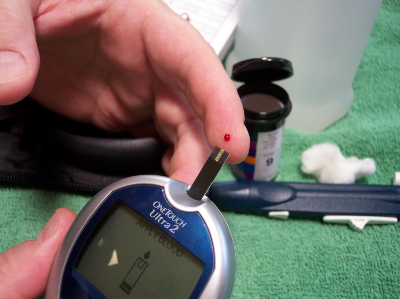
Type 1 diabetes (T1D) is a disease in which your immune system attacks and destroys your insulin-producing cells.  There is a known genetic risk factor in developing T1D, but there are also significant non-genetic components to getting the disease.  Previous research in mice has established the microbiome's connection with the development of diabetes, but the link in humans has not been studied as closely.  Researchers from various institutions in the U.S. and Finland recently assembled a cohort of infants genetically at-risk for diabetes, and tracked the changes in their microbiomes.  They discovered that the microbiomes of those individuals that were eventually diagnosed with diabetes underwent characteristic shifts leading up to diagnosis, and that these changes were not observed in healthy infants.  They published the results of their study in Cell Host and Microbe.  

The researchers sampled the stools of 33 infants in Finland and Estonia that were genetically at-risk for diabetes.  Their first major discovery was that even though the bacterial composition of the microbiome grew, changed, and became more diverse with age, the types and number of genetic pathways that were expressed by the microbiome, as well as the metabolites produced by the microbiome remained stable.  They also found many similar bacterial species between infants, however these infants usually had different strains of said bacterial species.  In most of these cases, once a particular strain established itself in the gut it remained stable and would not be displaced.

The scientists tracked the microbiome changes that occurred with diet as well.  During breast feeding Bifidobacterium and lactobacillus predominated, and Lachnospiraceae decreased.  After cessation of breast feeding the addition of eggs barley and soy seemed to have a direct influence on the microbiome.  One of the biggest factors in the developing microbiome was actually geography, as the Estonian infants had significantly higher levels of Bacteroides and Streptococcus species.

The researchers then compared the microbiome samples between those infants that were eventually diagnosed with diabetes and those that were not.  They discovered that a few bacterial species were much more abundant in those infants that got diabetes: Blautia, the Rikenellaceae, and the Ruminococcus and Streptococcus genera, including Ruminococcus gnavus and Streptococcus infantarius.  Interestingly, each of these bacteria are ‘pathobionts’, or bacteria which exist in many healthy peoples’ microbiomes but have the potential to become pathogenic.  Also, certain bacteria such as Coprococcus eutactus and Dialister invisus were non-existent in the diabetics' guts.  In addition, the researchers discovered that the expression of specific genes, like those associated with sugar transport and the biosynthesis of amino acids, underwent shifts prior to the onset of diabetes.  Finally, many of these bacteria that were associated with T1D appeared right before the onset of the disease, and these bacteria were linked to the presence and absence of certain metabolites in the stool.

These results provide exhaustive evidence for an association between the microbiome and diabetes.  It links specific bacteria in the microbiome and the expression of certain genes by the microbiome to the disease.  The next step is to study the mechanisms by which the microbiome induces diabetes, and then therapeutics can be developed.


Please email blog@MicrobiomeInstitute.org for any comments, news, or ideas for new blog posts.

The views expressed in the blog are solely those of the author of the blog and not necessarily the American Microbiome Institute or any of our scientists, sponsors, donors, or affiliates.