
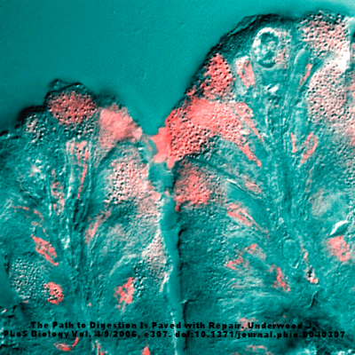
A surface mucous cell bordering on the stomach lumen secretes mucus (pink stain).
Our diet is full of various carbohydrates, composed of different monosaccharides and polysaccharides. Many of these survive our own digestion and make it all the way to the colon where they modulate our microbiome. Another source of saccharides for our gut bacteria is the mucous that we produce, which can be a rich source of fucose or sialic acid. Sialic acid has been implicated in many inflammatory diseases, such as bacterial vaginosis. Last week, researchers from Switzerland showed that sialic acid may play a critical role in colitis, at least in one colitis model commonly used in mice. They published their results in Nature Communications.
One way to induce intestinal inflammation in mice is to feed them dextran sodium sulfate (DSS). The reason this molecule causes colitis in this mice is unknown, but it is used in many models of the disease. In order to understand the possible role of sialic acid in the colitis, scientists created mice that could not produce mucous with sialic acid. They quickly realized that these mice were not as susceptible to the DSS-induced colitis as their normal counterparts. After, they tested how various antibiotics affected colitis in the DSS—colitis mice and discovered that Escherichia coli abundance was directly associated with the severity of the disease. Putting these ideas together, they tested and discovered that E. coli used sialic acid as their main carbohydrate source in vitro.
Interestingly, the E. coli cannot actually access the sialic acid from mucins, but instead need other bacteria, such as Bacteroides vulgatus to cleave and release the sialic acid from the mucins in order to access it. If sialic acid is indeed important to the human form of the disease there may be multiple approaches to combatting the disease. First, by eliminating E. coli, and second by eliminating the free sialic acid.


