Salmonella

The gut-vascular barrier’s role in regulating bacterial systemic dissemination

Microbiota that populate the gut do not have access to lymphatics, the liver, or other peripheral tissues in healthy individuals.  The gut-vascular barrier (GVB) plays an essential role in limiting bacteria access to systemic circulation in addition to protecting against pathogenic invasion.  However, some pathogenic bacteria can reach these tissues and induce a systemic immune response, but how this is exactly occurs is unknown.  Researchers from Italy sought to address this by exploring how GVB disruption lends itself to bacterial-pathogenesis in liver and lymphatic tissue sites. 

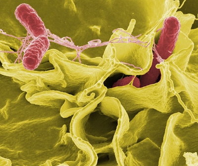
Two groups of mice were analyzed, one healthy control group and one group infected with Salmonella, a bacteria selected to study the pathogenic effects on the GVB.  Healthy control mice were first analyzed using fluorescent dextran polymer dyes at different sizes to assess the leakage of the dye into the intestines for visualization and characterization of the GVB.  After injection, intestinal blood vessels were analyzed using two-photon microscopy, and it was observed that an endothelial barrier could discriminate particles that are functionally similar according to size.  Specifically, dextran polymers 4 kilodaltons (kD) in size could enter the blood stream freely, while dextran polymers 70 kD in size could not.  The same experiment was then repeated in mice who were administered Salmonella prior to dextran, and interestingly the 70 kD polymers diffused freely into the blood stream, suggesting that the Salmonella disrupted the GVB.  Exploring deeper, the experimenters characterized the endothelial cell types that formed this tight-junction barrier, as it was found that plasma-lemma vesicle-associated protein-1 (PV1) was up-regulated in blood 6 hours after being exposed to Salmonella.  Furthermore, this finding was associated with systemic damage present in other organs. 

In another experiment using the dextran dye, the researchers determined that the dye reached the liver in mice infected with Salmonella after 60 minutes following injection, as it was concluded that the Salmonella traveled to the liver via portal vein circulation as opposed to lymphatic transport (where it would be present in the spleen).  Lastly, it was determined that Salmonella could discretely regulate B-catenin signaling by blocking these signals from being released in vascular endothelial cells. 

These findings highlight important mechanistic features of a physiological construct potentially implicated in many pathogenic diseases.  In the context of liver damage and celiac disease, these findings could link the GVB as drivers of pathogenesis as the GVB is disrupted in celiac patients.  Disruptions to the GVB deserve more investigation as this physiological barrier could be implicated in many pathogenic diseases.  

Please email blog@MicrobiomeInstitute.org for any comments, news, or ideas for new blog posts.

The views expressed in the blog are solely those of the author of the blog and not necessarily the American Microbiome Institute or any of our scientists, sponsors, donors, or affiliates.

Can gut microbes be used to diagnose and treat malnutrition?

Credit: Tanya Yatsunenko

Credit: Tanya Yatsunenko

When people think about malnutrition, they often think that not eating enough food leads to stunted growth, neurocognitive issues, weakened immune systems, and other health problems associated with malnutrition. While this is largely true, food scarcity and insecurity does lead to undernutrition, it is not the sole contributing factor to this pervasive global health problem.  Jeffrey Gordon and his group at Washington University School of Medicine in St. Louis have shown once again that gut microbes play an important role in undernutrition in a paper in Science Translational Medicine

To show the importance of the microbiome in undernutrition, Gordon’s team studied children in Malawi who were undernourished and others that were not. Specifically, they studied individuals with kwashiorkor, a form of severe undernutrition that occurs in children who often eat similar diets as other healthy children. They studied identical twins, one with the disease and one without the disease and sampled their gut microbes.  They transplanted the bacteria from the sick child into germ-free mice to see what effects the bacteria would have. When transplanted into the mice, the bacteria were very harmful causing weight loss as well as severe damage to the lining of the intestines and colon.

The scientists looked for bacteria that were targeted by an important molecule of the immune system called immunoglobin A (IgA). IgA is prevalent throughout the body and specifically in the gut. It plays an important role in preventing the bacteria in the gut from interacting with the human cells that line our intestines. As we saw in the paper on the blog on Monday about emulsifiers in our food, when gut bacteria in the gut interacts with the epithelial cells of the gut lining, severe health problems can arise. The scientists found that IgA and the immune system largely targeted Enterobacteriaceae, a large family of bacteria found in the gut that includes E. coli, Salmonella, and many others. The scientists were able to prevent weight loss in the mice by transplanting two strains of IgA targeted bacteria from the guts of healthy children into the mice, before they were exposed to the bacteria from the undernourished child.

This is an important study as it not only shows the significant role that gut bacteria have on malnutrition, but it shows that it may be possible to use the microbiome as a diagnostic tool to identify which children are at risk for undernutrition, and it may also be a therapeutic target for intervention. The scientists also studied 19 other groups of twins and found that higher levels of Enterobacteriaceae led to a greater risk of kwashiorkor. By sampling children at a very early age for gut bacteria, it could be possible to identify which children were at greater risk of becoming malnourished and intervening with probiotics or other therapeutic foods to alter the microbiome.

Please email blog@MicrobiomeInstitute.org for any comments, news, or ideas for new blog posts.

The views expressed in the blog are solely those of the author of the blog and not necessarily the American Microbiome Institute or any of our scientists, sponsors, donors, or affiliates.

Major advance in possible bacterial treatment for cancers

Salmonella bacteria

Salmonella bacteria

Editor’s note: Yesterday we wrote about how microbiome bacteria may be protecting cancer cells, but today we wanted to write about how microbiome bacteria can be engineered to kill cancer cells.  Enjoy.

In 2003 a group at the Harvard Medical School discovered that nonpathogenic Salmonella (a microbiome bacterium) that was injected into mice would preferentially accumulate in the tumors of those mice, sometimes by a factor of 10,000.  Soon after, the researchers tried to apply these findings to target and destroy cancer cells.  They began incorporating cancer-fighting proteins into the Salmonella with the hopes that the bacteria would accumulate in tumors and destroy them.  This was effective in killing the cancer, but the Salmonella was not specific enough to tumors, and the low levels that existed in healthy tissues still expressed anticancer proteins which killed the healthy tissues.  Recently though, this problem may have been solved.  One of the original scientists from Harvard Med, who now has his own group at UMass Amherst, developed a clever way to only trigger the anticancer proteins in Salmonella that are on cancer cells.  His group published their results in the Proceedings of the National Academy of Sciences on Wednesday.

The scientists incorporated a genetic switch in the Salmonella which would only trigger the production of anticancer proteins around cancer cells.  In order to do this they took advantage of the fact that the Salmonella accumulates to higher concentrations on cancer cells.  Many bacteria have proteins called quorum sensing proteins.  They are used by individuals and communities to sense what is around them, and to communicate with other bacteria.  Some of these quorum sensing proteins are only activated by their genes when there are enough other bacteria around them.  The scientists from UMass utilized this fact to incorporate a quorum sensing gene into Salmonella that would only activate a specific protein when it was around a high concentration of other Salmonella (e.g. in cancer cells). 

The scientists incorporated this quorum sensing gene into Salmonella so that, when triggered, it would express a fluorescent protein (which could be easily visualized).  They then injected these Salmonella into mice with various tumors.  They discovered, as they had hoped, that the fluorescing protein was predominantly expressed in cancer cells, and at very low levels elsewhere.  Moreover, the fluorescent protein was expressed for at least 24 days, and it did not appear to be expressed in other tissues (such as the liver) at all.

These experiments provide a partial proof of concept for a unique bacterial treatment to cancer.  The next step is to test anticancer proteins instead of fluorescing proteins, but the results using the fluorescing proteins are promising.  The scientists mentioned that the Salmonella used is non-pathogenic and can be eliminated from the body through natural processes.  This is a rather innovative potential cancer treatment, and we are excited to see what its future holds.

Please email blog@MicrobiomeInstitute.org for any comments, news, or ideas for new blog posts.

The views expressed in the blog are solely those of the author of the blog and not necessarily the American Microbiome Institute or any of our scientists, sponsors, donors, or affiliates.