ruminococcus gnavus

A new way in which some gut bacteria rely on their hosts’ mucous for energy

It is often understood that our gut bacteria live off of the foods we eat.  However, many gut bacteria can actually metabolize the mucous that protects the lining of our gut.  In fact, many bugs have the ability to digest a specific sugar that is attached to our mucins, called sialic acid.  Interestingly, some bacteria have the ability to cleave this sugar from the mucins, others have the ability to consume this sugar once it is released, and others have the ability to perform both of these tasks.  Last week, a new method for how gut bacteria can transform sialic acid was discovered, that some bacteria can actually transform sialic acid before cleaving it, and that this may be clinically relevant for Crohn’s disease and colitis.  The authors published their results in Nature Communications.

The authors were testing a common commensal bacteria, Ruminococcus gnavus, and noted that it had the ability to both cleave and consume sialic acid from gut mucins.  When they identified the metabolites from this process they discovered that the sialic acid was actually being converted to a different form by these bugs.  After further experimentation they realized that a type of enzyme, called an intramolecular trans sialidase, which had never before been observed in gut bacteria, was responsible.  The researchers then compared the genes from R. gnavus to other bugs common in the gut and noted that a full 11% of human gut commensals had this enzyme, and that these bacteria were overrepresented in people with IBD.  The authors think that the bugs who code for this enzyme have an inherent advantage over other gut microbiota because after they transform the sialic acid they can still use it for energy, whereas other bugs cannot, leaving the sugar all to themselves. 

The paper did not discuss specific mechanisms as to why these bugs may be overrepresented in Crohn’s and colitis.  They did however test a few molecules that inhibited the activity of the enzymes.  Perhaps if these enzymes or the responsible bugs are the cause of IBD, than these inhibitors could be used as therapeutics to combat the disease. 

Please email blog@MicrobiomeInstitute.org for any comments, news, or ideas for new blog posts.

The views expressed in the blog are solely those of the author of the blog and not necessarily the American Microbiome Institute or any of our scientists, sponsors, donors, or affiliates.

Emulsifiers in food cause many adverse health effects in mice

Emulsifiers are often used in ice cream to keep it stable and to give it it's texture.

Emulsifiers are often used in ice cream to keep it stable and to give it it's texture.

People often have spirited and impassioned views on the safety and consequences of adding ‘unnatural’ molecules to food.  An important aspect of this debate that everyone must keep in mind is the impossibility of testing every component of every molecule for its safety and long term impacts.  That being said, it should come as no surprise that new research can often teach us about the unexpected and overlooked safety issues regarding many food additives.  The newest class of compounds to come under scrutiny is emulsifiers, and a new paper published in Nature last week shows that these compounds may negatively impact the body through modulation of the intestinal lining and the microbiome. 

Emulsifiers are compounds that increase the stability of an emulsion.  They are often molecules like surfactants that have two parts, hydrophobic carbon chains and hydrophilic polar head groups.  Soap and egg yolks are common examples of emulsifiers.  There  are, of course, chemically produced emulsifiers as well that are often used in food.  Two examples of these, which were the emulsifying compounds used in the study, are polysorbate-80 (P80) and carboxymethylcellulose (CMC), and they are added to all sorts of foods like ice cream and pudding.  Evidence from this paper suggests though, that at least in mice these emulsifiers are wreaking havoc on the gut and microbiome.

A team of researchers from Israel, Cornell, and Emory did a variety of experiments on mice that were fed either of these emulsifiers in their water (at a concentration of 1%, similar to the levels added to human food.)  They first noticed that these mice had greatly compromised mucous layers on their gut, which allowed for bacteria to actually reach and be in contact with their epithelial gut cells.  In these mice gut permeability (leaky gut), inflammation, and incidences of colitis were all increased.  In addition, the inflammatory response and gut permeability were directly related to the average distance of bacteria to the actual epithelial layer; the closer the bacteria the more inflammation and permeability.

The researchers also measured the microbial populations of the feces in these mice and those that were eating emulsifiers had much less diverse microbiomes, which were enriched in Proteobacter, which are known to be associated with inflamed guts, and reduced in Bacteroidales, which are associated with healthy guts.  Also those eating emulsifiers had in increase in Ruminococcus gnavus which is associated with type 1 diabetes, as we have written about in the past. Interestingly, those mice that were given the emulsifiers tended to eat a lot more food than there control counterparts, and this led to weight gain and obesity amongst the mice drinking emulsifiers.  Moreover, these same mice had higher fasting glucose levels, indicative of impaired glycemic control and metabolic syndrome.  The scientists tested if these effects were seen in mice that were fed the emulsifiers in their food, rather than in their water, and the same outcomes were observed.  In addition, the scientists observed shifts in the production of certain short chained fatty acids and bile acids produced by the microbiome in mice fed emulsifiers (click on the tags below to read about the wide range of health effects that both these compounds have been implicated with).

The researchers then did a series of experiments that showed that it was the actual shifts in the microbiome populations and not just the change in mucous that was primarily responsible for the adverse health effects in the mice given emulsifiers.  For example, germ free mice that were given  emulsifiers did not have compromised mucous, and many of the negative health effects like inflammation were not observed.  On the other hand, performing a microbiome transplant from a mouse given the emulsifiers to a mouse that was not given the emulsifiers did result in these negative health effects, including the bacterial penetration of mucous to the epithelial lining.

This was a fantastic article that may, in time, prove to be immensely important.  Of course all the usual caveats apply, such as studies in mice are not indicative of human responses, and more studies must be performed in order to confirm these findings.  Still though, the introduction of emulsifiers into the mice’s diets resulted in many of the negative health impacts that are associated with the microbiome, something that we really haven’t seen in the literature before now.

Please email blog@MicrobiomeInstitute.org for any comments, news, or ideas for new blog posts.

The views expressed in the blog are solely those of the author of the blog and not necessarily the American Microbiome Institute or any of our scientists, sponsors, donors, or affiliates.

The infant microbiome changes before the onset of type 1 diabetes

file0001403630450.jpg

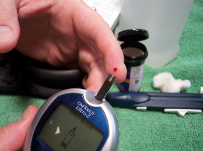
Type 1 diabetes (T1D) is a disease in which your immune system attacks and destroys your insulin-producing cells.  There is a known genetic risk factor in developing T1D, but there are also significant non-genetic components to getting the disease.  Previous research in mice has established the microbiome's connection with the development of diabetes, but the link in humans has not been studied as closely.  Researchers from various institutions in the U.S. and Finland recently assembled a cohort of infants genetically at-risk for diabetes, and tracked the changes in their microbiomes.  They discovered that the microbiomes of those individuals that were eventually diagnosed with diabetes underwent characteristic shifts leading up to diagnosis, and that these changes were not observed in healthy infants.  They published the results of their study in Cell Host and Microbe.  

The researchers sampled the stools of 33 infants in Finland and Estonia that were genetically at-risk for diabetes.  Their first major discovery was that even though the bacterial composition of the microbiome grew, changed, and became more diverse with age, the types and number of genetic pathways that were expressed by the microbiome, as well as the metabolites produced by the microbiome remained stable.  They also found many similar bacterial species between infants, however these infants usually had different strains of said bacterial species.  In most of these cases, once a particular strain established itself in the gut it remained stable and would not be displaced.

The scientists tracked the microbiome changes that occurred with diet as well.  During breast feeding Bifidobacterium and lactobacillus predominated, and Lachnospiraceae decreased.  After cessation of breast feeding the addition of eggs barley and soy seemed to have a direct influence on the microbiome.  One of the biggest factors in the developing microbiome was actually geography, as the Estonian infants had significantly higher levels of Bacteroides and Streptococcus species.

The researchers then compared the microbiome samples between those infants that were eventually diagnosed with diabetes and those that were not.  They discovered that a few bacterial species were much more abundant in those infants that got diabetes: Blautia, the Rikenellaceae, and the Ruminococcus and Streptococcus genera, including Ruminococcus gnavus and Streptococcus infantarius.  Interestingly, each of these bacteria are ‘pathobionts’, or bacteria which exist in many healthy peoples’ microbiomes but have the potential to become pathogenic.  Also, certain bacteria such as Coprococcus eutactus and Dialister invisus were non-existent in the diabetics' guts.  In addition, the researchers discovered that the expression of specific genes, like those associated with sugar transport and the biosynthesis of amino acids, underwent shifts prior to the onset of diabetes.  Finally, many of these bacteria that were associated with T1D appeared right before the onset of the disease, and these bacteria were linked to the presence and absence of certain metabolites in the stool.

These results provide exhaustive evidence for an association between the microbiome and diabetes.  It links specific bacteria in the microbiome and the expression of certain genes by the microbiome to the disease.  The next step is to study the mechanisms by which the microbiome induces diabetes, and then therapeutics can be developed.


Please email blog@MicrobiomeInstitute.org for any comments, news, or ideas for new blog posts.

The views expressed in the blog are solely those of the author of the blog and not necessarily the American Microbiome Institute or any of our scientists, sponsors, donors, or affiliates.